无机材料学报 ›› 2013, Vol. 28 ›› Issue (1): 21-28.DOI: 10.3724/SP.J.1077.2013.12269 CSTR: 32189.14.SP.J.1077.2013.12269
吕晓迎, 黄 炎, 俞亚东, 杨雅敏
收稿日期:2012-04-26
修回日期:2012-07-18
出版日期:2013-01-10
网络出版日期:2012-12-20
基金资助:LÜ Xiao-Ying, HUANG Yan, YU Ya-Dong, YANG Ya-Min
Received:2012-04-26
Revised:2012-07-18
Published:2013-01-10
Online:2012-12-20
Supported by:摘要:
生物材料的生物相容性是生物材料研究领域的关键科学问题。分子生物学技术的发展使生物材料的生物相容性评价从动物水平和细胞水平深入到分子水平。生物组学技术的发展为高通量进行生物材料分子生物相容性评价、阐明生物材料与生物体的相互作用机理提供了有效的手段。本文综述了生物材料生物相容性研究现状及基因组学、蛋白质组学技术在生物材料生物相容性研究中的应用。
中图分类号:
吕晓迎, 黄 炎, 俞亚东, 杨雅敏. 基因/蛋白质组学技术在生物材料生物相容性研究中的应用[J]. 无机材料学报, 2013, 28(1): 21-28.
Lü Xiao-Ying, HUANG Yan, YU Ya-Dong, YANG Ya-Min. Application of Genomics/Proteomics Technologies in the Research of Biocompatibility of Biomaterials[J]. Journal of Inorganic Materials, 2013, 28(1): 21-28.
| Definition | Source | |
|---|---|---|
| 1986 | Biocompatibility is the capability of a prosthesis implanted in the body to exist in harmony with tissue without causing deleterious changes. | International Dictionary of Medicine and Biology |
| 1987 | Biocompatibility refers to the ability of a material to perform with an appropriate host response in a specific situation. | Williams D F[ |
| 2003 | The quality of not having toxic or injurious effects on biological systems. | Dorland's Medical Dictionary |
| 2006 | ‘‘Biocompatibility’’ is defined not only by the lack of cytotoxicity of a biomaterial, but also by the biofunctionality of the material, which enables it to support cell-biomaterial interactions according to the local and organ-specific situation where the biomaterial is applied. | Rickert D[ |
| 2008 | Comparison of the tissue response produced through the close association of the implanted candidate material to its implant site within the host animal to that tissue response recognized and established as suitable with control materials. | American Society for Testing and Materials (ASTM) |
| 2008 | Biocompatibility refers to the ability of a biomaterial to perform its desired function with respect to a medical therapy, without eliciting any undesirable local or systemic effects in the recipient or beneficiary of that therapy, but generating the most appropriate beneficial cellular or tissue response in that specific situation, and optimising the clinically relevant performance of that therapy. | Williams D F[ |
Table 1 Definitions of biocompatibility
| Definition | Source | |
|---|---|---|
| 1986 | Biocompatibility is the capability of a prosthesis implanted in the body to exist in harmony with tissue without causing deleterious changes. | International Dictionary of Medicine and Biology |
| 1987 | Biocompatibility refers to the ability of a material to perform with an appropriate host response in a specific situation. | Williams D F[ |
| 2003 | The quality of not having toxic or injurious effects on biological systems. | Dorland's Medical Dictionary |
| 2006 | ‘‘Biocompatibility’’ is defined not only by the lack of cytotoxicity of a biomaterial, but also by the biofunctionality of the material, which enables it to support cell-biomaterial interactions according to the local and organ-specific situation where the biomaterial is applied. | Rickert D[ |
| 2008 | Comparison of the tissue response produced through the close association of the implanted candidate material to its implant site within the host animal to that tissue response recognized and established as suitable with control materials. | American Society for Testing and Materials (ASTM) |
| 2008 | Biocompatibility refers to the ability of a biomaterial to perform its desired function with respect to a medical therapy, without eliciting any undesirable local or systemic effects in the recipient or beneficiary of that therapy, but generating the most appropriate beneficial cellular or tissue response in that specific situation, and optimising the clinically relevant performance of that therapy. | Williams D F[ |
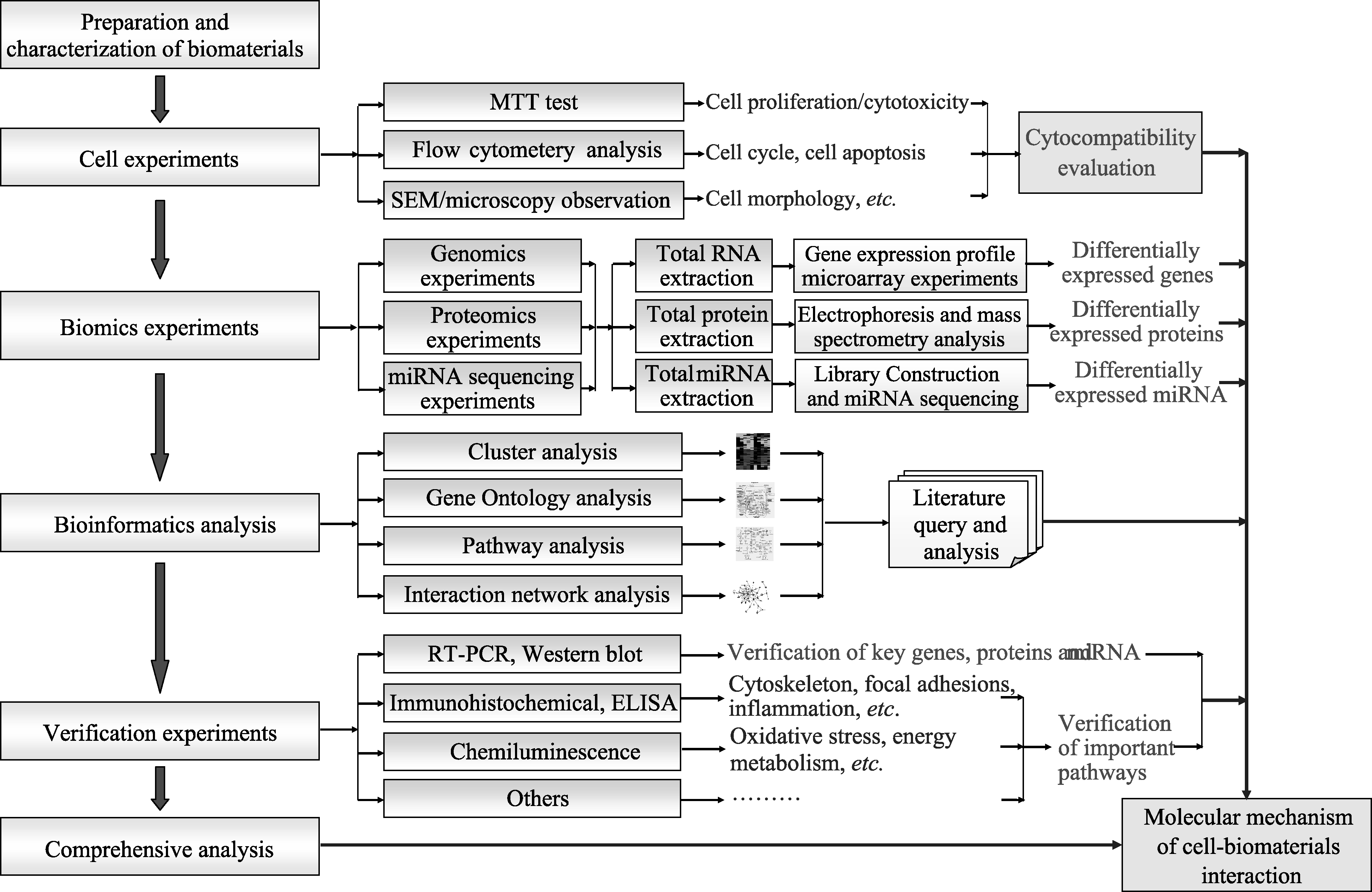
图1 基于生物组学和生物信息学技术的生物材料分子生物相容性研究技术路线
Fig. 1 Common routes of the molecular biocompatibilty study based on biomics and bioinformatics technologies
| [1] | Hegyeli R J. Limitations of current techniques for assessing biohazards and biocompatibility of new candidate materials-orpl. Abstracts of papers of the American Chemical Society, 1970, 1. |
| [2] | Homsy C A, Ansevin KD, Obannon W, et al. Rapid in-vitro screening of polymers for biocompatibility. J. Macromol. Sci. A, 1970, 4(3): 615. |
| [3] | 顾汉卿,徐国风. 生物医学材料学. 天津: 天津科技翻译出版公司, 1993: 92. |
| [4] | Williams D F. Definitions in biomaterials. Progress in biomedical engineering, Amsterdam: Elsevier, 1987: 72. |
| [5] | Rickert D, Lendlein A, Peters I, et al. Biocompatibility testing of novel multifunctional polymeric biomaterials for tissue engineering applications in head and neck surgery: an overview. Eur. Arch. Otorhinolaryngol., 2006, 263(3): 215-222. |
| [6] | Williams D F. On the mechanisms of biocompatibility. Biomaterials, 2008, 29(20): 2941-2953. |
| [7] | Lü XY, Bao X, Huang Y, et al. Mechanisms of cytotoxicity of nickel ions based on gene expression profiles. Biomaterials, 2009, 30(2): 141-148. |
| [8] | Chou L S, Firth J D, Uitto V J, et al. Substratum surface topog- raphy alters cell shape and regulates fibronectin mRNA level, mRNA stability, secretion and assembly in human fibroblasts. J. Cell Sci., 1995, 108(4): 1563-1573. |
| [9] | Chou L S, Firth J D, Nathanson D, et al. Effects of titanium on transcriptional and post-transcriptional regulation of fibronectin inhuman fibroblasts. J. Biomed. Mater. Res., 1996, 31(2): 209-217. |
| [10] | Chou L S, Firth J D, Uitto V J, et al. Effects of titanium substratum and grooved surface topograhy on metalloproteinase-2 expression in human fibroblasts. J. Biomed. Mater. Res., 1998, 39(3): 437-445. |
| [11] | Ripamonti U, Crooks J, Khoah L, et al. The induction of bone formation by coral-derived calcium carbonate/hydroxyapatite constructs. Biomaterials, 2009, 30(7): 1428-1439. |
| [12] | Park S H, Gil E S, Shi H, et al. Relationships between degradability of silk scaffolds and osteogenesis. Biomaterials, 2010, 31(24): 6162-6172. |
| [13] | Im D D, Kruger E A, Huang W B R, et al. Extracellular-signal- related kinase 1/2 is responsible for inhibition of osteogenesis in three-dimensional cultured MC3T3-E1 cells. Tissue Eng. A, 2010, 16(11): 3485-3494. |
| [14] | Brown P O, Botstein D. Exploring the new world of the genome with DNA microarrays. Nat. Genet.(Suppl), 1999, 21(1): 33-37. |
| [15] | Peng J, Gygi S P. Proteomics: the move to mixtures. J. Mass Spectrom,. 2001, 36(10): 1083-1091. |
| [16] | Yuan Q, Zhao F K. New frontiers in the proteome research: quantitative proteomics. Acta Biochimica Et Biophysica Sinica., 2001, 33(5): 477-482. |
| [17] | Mueller P P, May T, Perz A, et al. Control of muscle cell proliferation by ferrous iron. Biomaterials, 2006, 27(10): 2193-2200. |
| [18] | Lü X Y, Lu H Q, Zhao L F, et al. Genome-wide pathways analysis of nickel ion-induced differential genes expression in fibroblasts. Biomaterials, 2010, 31(8): 1965-1973. |
| [19] | Kasprzak K S, Sunderman F W, Salnikowa K. Nickel carcinogenesis. Mutat. Res.-Fund. Mol. M., 2003, 533(1/2): 67-97. |
| [20] | Zhao L F, Hong Y, Yang D Y, et al. The underlying biological mechanisms of biocompatibility differences between bare and TiN-coated NiTi alloys. Biomed. Mater. , 2011, 6(2): 025012-1-12. |
| [21] | Hong Y, Zhao L F, Yang D Y, et al. Analyses of differentially expressed genes in human cell in response to uncoated and Ti-coated NiTi alloys. The 2011 International Conference on Information Science and Technology (ICIST 2011), Nanjing, 2011: 1377-1381. |
| [22] | Yang D Y, Hong Y, Zhao L F, et al. Proteomic analysis of human endothelial cells in response to bare and titanium nitride-coated NiTi alloys. 3rd Chinese-Europe Symposium on Biomaterials in Regenerative Medicine, Nanjing, 2011: 159. |
| [23] | Lee H, Ko S H. Microarray analysis of MC3T3-E1 osteoblastic cell response to machined titanium surface and resorbable blast material titanium surface. Bone, 2011, 48(Suppl 2): S111. |
| [24] | Sollazzo V, Palmieri A, Pezzetti F, et al. Genetic effect of zirconium oxide coating on osteoblast-like cells. J. Biomed. Mater. Res. B, 2008, 84B(2): 550-558. |
| [25] | Derhami K, Zheng J, Li L, et al. Proteomic analysis of human skin fibroblasts grown on titanium: novel approach to study molecular biocompatibility. J. Biomed Mater Res., 2001, 56(2): 234-244. |
| [26] | Leven R M, Virdi A S, Sumner D R. Patterns of gene expression in rat bone marrow stromal cells cultured on titanium alloy discs of different roughness. J. Biomed. Mater. Res. A, 2004, 70A(3): 391-401. |
| [27] | Zhao M, An M, Wang Q, et al. Quantitative proteomic analysis of human osteoblast-like MG-63 cells in response to bioinert implant material titanium and polyetheretherketone,J. Proteomics, 2012, 75(12): 3560-3573. |
| [28] | Xie J, Baumann M J, McCabe L R. Osteoblasts respond to hydroxyapatite surfaces with immediate changes in gene expression. J. Biomed. Mater. Res., 2004, 71A(1): 108-117. |
| [29] | Song J H, Kim J H, Park S, et al. Signaling responses of osteoblast cells to hydroxyapatite: the activation of ERK and SOX9. J. Bone Miner. Metab., 2008, 26(2): 138-142. |
| [30] | Xu J L, Khor K A, Sui J J, et al. Comparative proteomics profile of osteoblasts cultured on dissimilar hydroxyapatite biomaterials: an iTRAQ-coupled 2-D LC-MS/MS analysis. Proteomics, 2008, 8(20): 4249-4258. |
| [31] | Xu J L, Khor K A, Sui J J, et al. Protein expression profiles in osteoblasts in response to differentially shaped hydroxyrapatite nanoparticles. Biomaterials, 2009, 30(29): 5385-5391. |
| [32] | 王健丹. 基于基因表达谱芯片技术的天然羟基磷灰石成骨诱导机理研究. 南京: 东南大学硕士论文, 2010. |
| [33] | Wang J D, Lü X Y, Li B, et al. Mechanism Study on Osteoinductive Property of Natural Hydroxyapatite to Bone Marrow Mesenchymal Stem Cell. National Conference on Biomaterials, Chengdu, 2010. |
| [34] | Zhang Z W, Lü X Y, Wang J D. Pilot Study of Osteoinduction mechanism of Natural Hydroxyapatite. 9th World Biomaterials Congress, Chengdu, 2012. |
| [35] | Carinci F, Piattelli A, Degidi M, et al. Genetic effects of anorganic bovine bone (Bio-Oss®) on osteoblast-like MG63 cells. Arch. Oral Biol., 2006, 51(2): 154-163. |
| [36] | Zhang L L, Hanagata N, Maeda M, et al. Porous hydroxyapatite and biphasic calcium phosphate ceramics promote ectopic osteoblast differentiation from mesenchymal stem cells. Sci. Tech. Adv. Mater., 2009, 10(2): 025003. |
| [37] | Khan J A, Pillai B, Das T K, et al. Molecular effects of uptake of gold nanoparticles in HeLa cells. ChemBioChem, 2007, 8(11): 1237-1240. |
| [38] | Kawata K, Osawa M, Okabe S. In vitro toxicity of silver nanoparticles at noncytotoxic doses to HepG2 human hepatoma cells. Environ. Sci. Technol., 2009, 43(15): 6046-6051. |
| [39] | Kim E, Chu Y C, Han J Y, et al. Proteomic analysis of silver nanoparticle toxicity in rat. Toxicol. Environ. Health. Sci., 2010, 2(4): 251-262. |
| [40] | Yang Y M, Qu Y H, LÜ X Y. Global gene expression analysis of the effects of gold nanoparticles on human dermal fibroblasts. J. Biomed Nanotechnol., 2010, 6(3): 234-246. |
| [41] | Ma J W, Lü X Y, Huang Y. Genomic analysis of cytotoxicity response to nanosilver in human dermal fibroblasts. J. Biomed Nanotechnol., 2011, 7(2): 263-275. |
| [42] | Lv X Y, Qu Y H, Yang Y M, et al. Proteomic analysis of molecular mechanism of interactions between gold nanoparticles and human dermal fibroblasts-fetal. Journal of Functional Materials, 2011, 42(6): 1016-1020. |
| [43] | Lü X Y, Huang Y, Ma J W. Mechanism study of cytotoxicity of silver nanoparticles based on biomics methods. Chinese Journal of Dental Materials and Devices, 2010, 19(4): 179-182. |
| [44] | Cui D X, Tian F R, Ozkan C S, et al. Effect of single wall carbon nanotubes on human HEK293 cells. Toxicol. Lett., 2005, 155(1): 73-85. |
| [45] | Ding L G, Stilwell J, Zhang T T, et al. Molecular characterization of the cytotoxic mechanism of multiwall carbon nanotubes and nano-onions on human skin fibroblast. Nano Lett., 2005, 5(12): 2448-2464. |
| [46] | Peng L, Barczak A J, Barbeau R A, et al. Whole genome expression analysis reveals differential effects of TiO2 nanotubes on vascular cells. Nano Lett., 2010, 10(1): 143-148. |
| [47] | Chew S Y, Mi R, Hoke A, et al. The effect of the alignment of electrospun fibrous scaffolds on Schwann cell maturation. Biomaterials, 2008, 29(6): 653-661. |
| [48] | Agudelo-Garcia P A, De Jesus J K, Williams S P, et al. Glioma cell migration on three-dimensional nanofiber scaffolds is regulated by substrate topography and abolished by inhibition of STAT3 signaling. Neoplasia, 2011, 13(9): 831-840. |
| [49] | He W, Yong T, Ma Z W, et al. Biodegradable polymer nanofiber mesh to maintain functions of endothelial cells. Tissue Eng., 2006, 12(9): 2457-2466. |
| [50] | Yu Y D, Lü X Y. Integrin roles in Aligned Nanofiber influence PC12 Cell Differentiation. 9th World Biomaterials Congress, Chengdu, 2012. |
| [51] | Dinnes D L M, Marcal H, Mahler S M, et al. Material surfaces affect the protein expression patterns of human macrophages: a proteomics approach. J. Biomed. Mater. Res. A, 2007, 80A(4): 895-908. |
| [52] | Rahman M A, Kumar S G, Kim S W, et al. Proteomic analysis for inhibitory effect of chitosan oligosaccharides on 3T3-L1 adipocyte differentiation. Proteomics, 2008, 8(3): 569-581. |
| [53] | Jaworski J, Klapperich C M. Fibroblast remodeling activity at two- and three-dimensional collagen-glycosaminoglycan interfaces. Biomaterials, 2006, 27(23): 4212-4220. |
| [54] | Klapperich C M, Bertozzi C R. Global gene expression of cells attached to a tissue engineering scaffold. Biomaterials, 2004, 25(25): 5631-5641. |
| [55] | Li G N, Livi L L, Gourd C M, et al. Genomic and morphological changes of neuroblastoma cells in response to three-dimensional matrices. Tissue Eng., 2007, 13(5): 1035-1047. |
| [56] | Xue M. Biological evaluation of medical devices-test animal welfare requirements. Chinese Journal of Dental Materials and Devices, 2007, 16(3): 117-124. |
| [57] | Johansson H, Lindstedt M, Albrekt A S, et al. A genomic biomarker signature can predict skin sensitizers using a cell-based in vitro alternative to animal tests. BMC Genomics, 2011, 12: 399-1-19. |
| [1] | 陈明俊, 缪洪康, 肖英俊, 邓建波, 张翔, 赵九蓬, 李垚. 光-热双响应材料研究进展: 从设计策略到智能窗应用[J]. 无机材料学报, 2026, 41(6): 723-738. |
| [2] | 宋坤洁, 解荣军. 机器学习驱动新型发光材料的研究进展[J]. 无机材料学报, 2026, 41(6): 689-703. |
| [3] | 胡钰晴, 朱一新, 乐先浩, 万青. 钽酸锂晶圆减薄技术及其热释电红外探测器应用进展[J]. 无机材料学报, 2026, 41(6): 764-774. |
| [4] | 刘春帆, 陈科, 葛芳芳, 黄庆. 核用耐铅铋腐蚀涂层的研究进展[J]. 无机材料学报, 2026, 41(6): 775-786. |
| [5] | 胡扬, 谢敏, 张筱怡, 李想, 郭新伟, 姜南, 周文瀚, 张胜利, 曾海波. 计算与数据驱动环保型发光材料的研究进展[J]. 无机材料学报, 2026, 41(6): 704-722. |
| [6] | 王俊卜, 黄泽皑, 杨茗凯, 蒙颖, 周明炜, 周莹. 甲烷转化用抗积碳催化材料研究进展[J]. 无机材料学报, 2026, 41(6): 739-750. |
| [7] | 王金文, 杨振, 周欢, 夏丹, 杨磊. 可注射无机材料及其生物医学应用[J]. 无机材料学报, 2026, 41(6): 751-763. |
| [8] | 李涵涛, 沈强, 罗国强, 王雪飞, 高明, 陈晨. 机械球磨法调控硅基负极材料结构与性能的研究进展[J]. 无机材料学报, 2026, 41(5): 561-572. |
| [9] | 解陈一, 缪花明, 张蔚然, 刘荣军, 王衍飞, 李端. 理论计算在高熵陶瓷领域的研究进展[J]. 无机材料学报, 2026, 41(5): 545-560. |
| [10] | 李璇, 叶奎材, 冯佳音, 邱家军, 钱文昊, 邢敏. 钛基牙种植体表面改性促进软组织封闭的研究进展[J]. 无机材料学报, 2026, 41(4): 432-444. |
| [11] | 彭德招, 李瑞, 王文鸿, 王梓瑞, 章志珍. 钠氯化物固态电解质研究进展[J]. 无机材料学报, 2026, 41(4): 409-420. |
| [12] | 陈坤, 姜勇刚, 冯军宗, 李良军, 胡艺洁, 冯坚. 锆酸镧多孔隔热材料研究进展[J]. 无机材料学报, 2026, 41(4): 421-431. |
| [13] | 韦连金, 齐志杰, 汪信, 朱俊武, 付永胜. 纳米金刚石改性及其在电催化氧还原反应中的应用[J]. 无机材料学报, 2026, 41(3): 273-288. |
| [14] | 刘占一, 李勉, 欧阳晓平, 柴之芳, 黄庆. 干法后处理熔盐中Sr/Cs去除方法的研究进展[J]. 无机材料学报, 2026, 41(2): 150-158. |
| [15] | 孙炼, 张磊磊, 薛泽旭, 吴坤, 陈晔, 李志远, 王鲁凯, 王尊刚. 面向辐射探测应用的零维金属卤化物闪烁体研究进展[J]. 无机材料学报, 2026, 41(2): 159-176. |
| 阅读次数 | ||||||
|
全文 |
|
|||||
|
摘要 |
|
|||||