Journal of Inorganic Materials ›› 2025, Vol. 40 ›› Issue (10): 1097-1110.DOI: 10.15541/jim20250025
Special Issue: 【信息功能】敏感陶瓷(202512)
• REVIEW • Previous Articles Next Articles
YUAN Long1( ), JIA Ru1, YUAN Meng1,2, ZHANG Jian2, DUAN Yu2, MENG Xiangdong1()
Received:2025-01-16
Revised:2025-02-18
Published:2025-10-20
Online:2025-05-09
Contact:
MENG Xiangdong, professor. E-mail: xdmeng@jlnu.edu.cnAbout author:YUAN Long (1988-), male, associate professor. E-mail: yuanlong@jlnu.edu.cn
Supported by:CLC Number:
YUAN Long, JIA Ru, YUAN Meng, ZHANG Jian, DUAN Yu, MENG Xiangdong. Mechanism and Application of X-ray Induced Photochromic Materials: A Review[J]. Journal of Inorganic Materials, 2025, 40(10): 1097-1110.
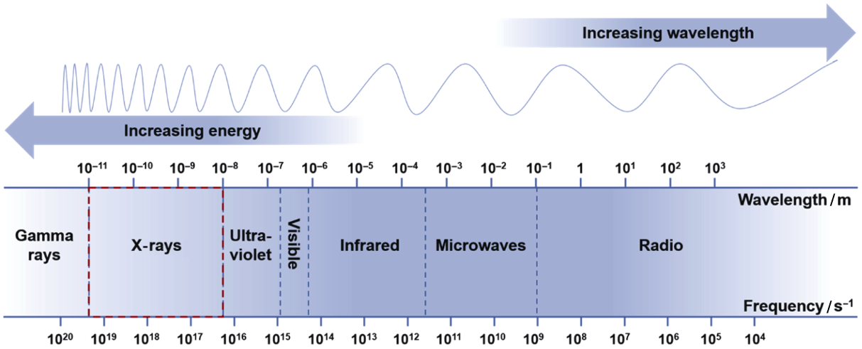
Fig. 1 Electromagnetic spectrum with the red box indicating the wavelength range corresponding to X-rays, and the curve above representing the electromagnetic wave energy
| Material system | Advantage | Disadvantage | Mechanism |
|---|---|---|---|
| Organic XP material (Iodine modified isomers based on 9,9'-(6-iodophenoxy- 1,3,5-triazine-2,4-diylbis (9h carbazole)[ compounds[ | Easy to regulate optical properties; Flexible, tunable photosensitive band and reaction rate through chemical modification; Lightweight, easy to shape, and suitable for various carriers | Poor stability and susceptibility to environmental factors such as humidity and oxygen; Low thermal stability | Reversible photoisomeri- zation or open-closed-loop reaction of organic molecules under X-ray excitation to achieve color change by adjusting the electronic transition energy level of the molecule |
| Organic inorganic hybrid XP material (Zn(II)-viologen coordination polymers[ | Combining flexibility of organic materials with stability of inorganic materials; Multifunctional and adjustable based on the ratio of organic and inorganic parts; Wide radiation response range tunable optical properties through material design | Complex synthesis, stability of interface bonding needs to be further improved; Complexity of material composites resulting in difficulty in mass production and repeatability control | Based on the interfacial charge transfer or energy transfer between organic and inorganic components, the synergistic initiation of structural reorganization or redox reaction leads to color change |
| Inorganic XP material (NaLuF4:Ho3+[ BaMgSiO4[ | High stability, high temperature resistance, and corrosion resistance; Strong responsiveness to high- energy radiation such as X-rays; Good thermal, light, and chemical stability, excellent long-term performance | Usually, less obvious color change than that of organic materials, difficult in controlling reversibility; Poor flexibility, difficult in processing into complex structures | X-rays induce lattice defects or metal ion valence state changes, changing the band structure of the material or forming a color center, and finally achieving color change |
Table 1 Advantages and disadvantages of XP materials[14-21]
| Material system | Advantage | Disadvantage | Mechanism |
|---|---|---|---|
| Organic XP material (Iodine modified isomers based on 9,9'-(6-iodophenoxy- 1,3,5-triazine-2,4-diylbis (9h carbazole)[ compounds[ | Easy to regulate optical properties; Flexible, tunable photosensitive band and reaction rate through chemical modification; Lightweight, easy to shape, and suitable for various carriers | Poor stability and susceptibility to environmental factors such as humidity and oxygen; Low thermal stability | Reversible photoisomeri- zation or open-closed-loop reaction of organic molecules under X-ray excitation to achieve color change by adjusting the electronic transition energy level of the molecule |
| Organic inorganic hybrid XP material (Zn(II)-viologen coordination polymers[ | Combining flexibility of organic materials with stability of inorganic materials; Multifunctional and adjustable based on the ratio of organic and inorganic parts; Wide radiation response range tunable optical properties through material design | Complex synthesis, stability of interface bonding needs to be further improved; Complexity of material composites resulting in difficulty in mass production and repeatability control | Based on the interfacial charge transfer or energy transfer between organic and inorganic components, the synergistic initiation of structural reorganization or redox reaction leads to color change |
| Inorganic XP material (NaLuF4:Ho3+[ BaMgSiO4[ | High stability, high temperature resistance, and corrosion resistance; Strong responsiveness to high- energy radiation such as X-rays; Good thermal, light, and chemical stability, excellent long-term performance | Usually, less obvious color change than that of organic materials, difficult in controlling reversibility; Poor flexibility, difficult in processing into complex structures | X-rays induce lattice defects or metal ion valence state changes, changing the band structure of the material or forming a color center, and finally achieving color change |
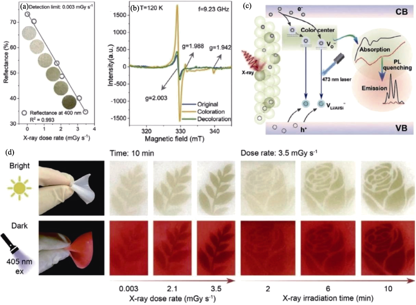
Fig. 2 Reversible formation mechanism of color center[45] (a) LAS-Sm relationship between diffuse reflectance intensity and X-ray dose rate accompanied by its corresponding photos; (b) EPR spectra of O element in initial state, photochromic state, and decolorized state; (c) Photochromic and luminescent modulation mechanism of LAS-Sm fluorescent powder; (d) Morphology of leaves and roses after irradiation with gradually prolonged time or different dose rates in bright and dark fields
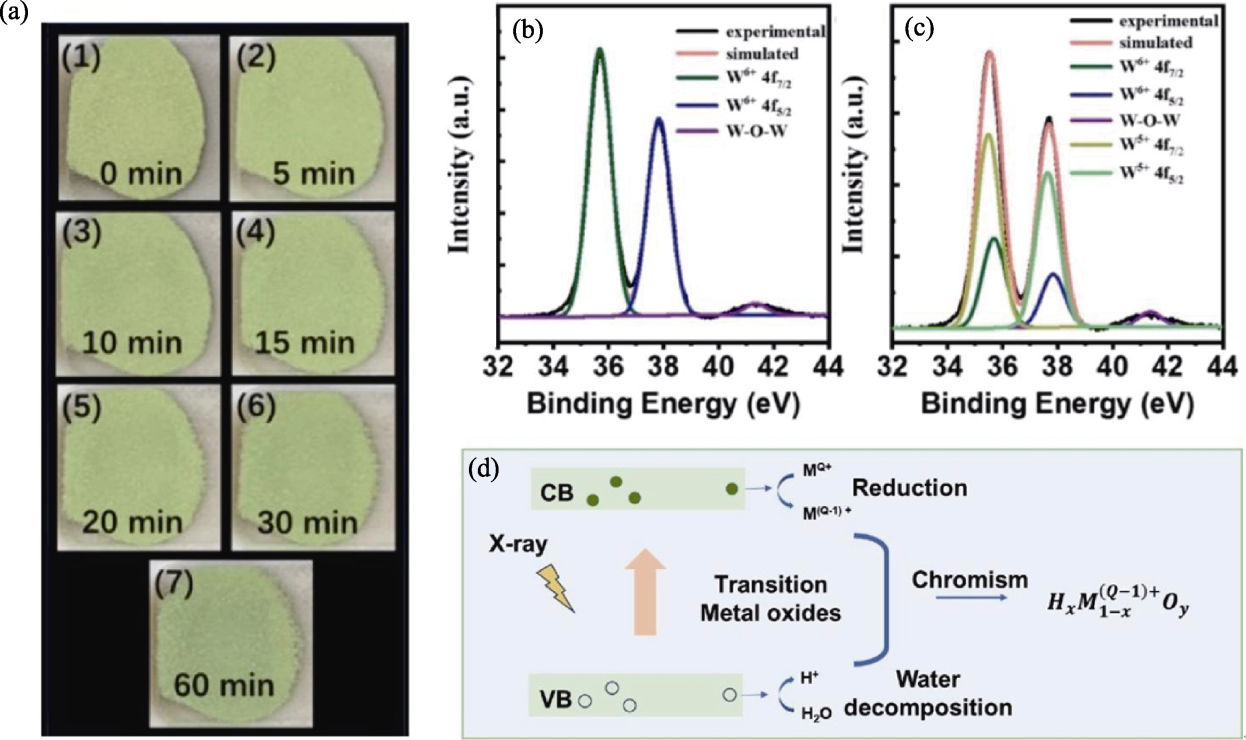
Fig. 3 Redox reaction mechanism[20,25] (a) Digital photos of WO3 powder under different X-ray irradiation times[20]; (b, c) XPS (Al-Kα) nuclear level spectra of WO3 powder before (b) and after (c) X-ray irradiation[20]; (d) General model for X-ray induced photochromism of transition metal oxides[25]
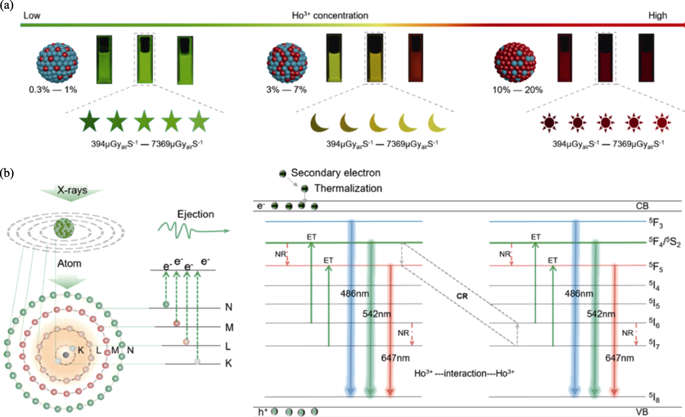
Fig. 4 Photoelectron transfer mechanism of defects associated with cross-relaxation[19] (a) Photos of NaLuF4: Ho3+ NCs doped with different concentrations of Ho3+ ions at different X-ray irradiation doses; (b) Color changing mechanism of NaLuF4: Ho3+ NCs
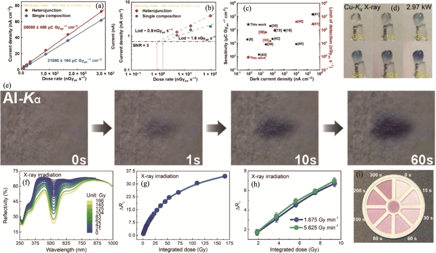
Fig. 5 Application of XP materials in X-ray detectors[18,21,27,57] (a-c) X-ray detection performance of single component and heterojunction detectors under 15 keV X-ray and -50 V bias[57]: (a) Current density of single component and heterojunction as a function of incident dose rate; (b) Dose dependent current density of single component and heterojunction with dashed line representing a signal-to-noise ratio (SNR) of 3; (c) Comparison of dark current density, sensitivity, and detection limit (LOD) of several advanced polycrystalline perovskite X-ray detectors; (d) Single crystal exposed to Cu-Kα X-ray (λ at 0.154056 nm, generator power at 2.97 kW) discoloration photo[18]; (e) Colour images of Al-Kα X-rays over time after 1 min of irradiation[27]; (f) Reflectance of BaMgSiO4 after exposure to different doses of X-rays (1.875 Gy·min-1)[21]; (g) Relationship between ΔR1 (coloring) and comprehensive X-ray irradiation dose[21]; (h) Dosimetric behavior of BaMgSiO4 under different X-ray irradiation intensities[21]; (i) Concept validation dosimeter for colorimetric detection of radiation dose[21]
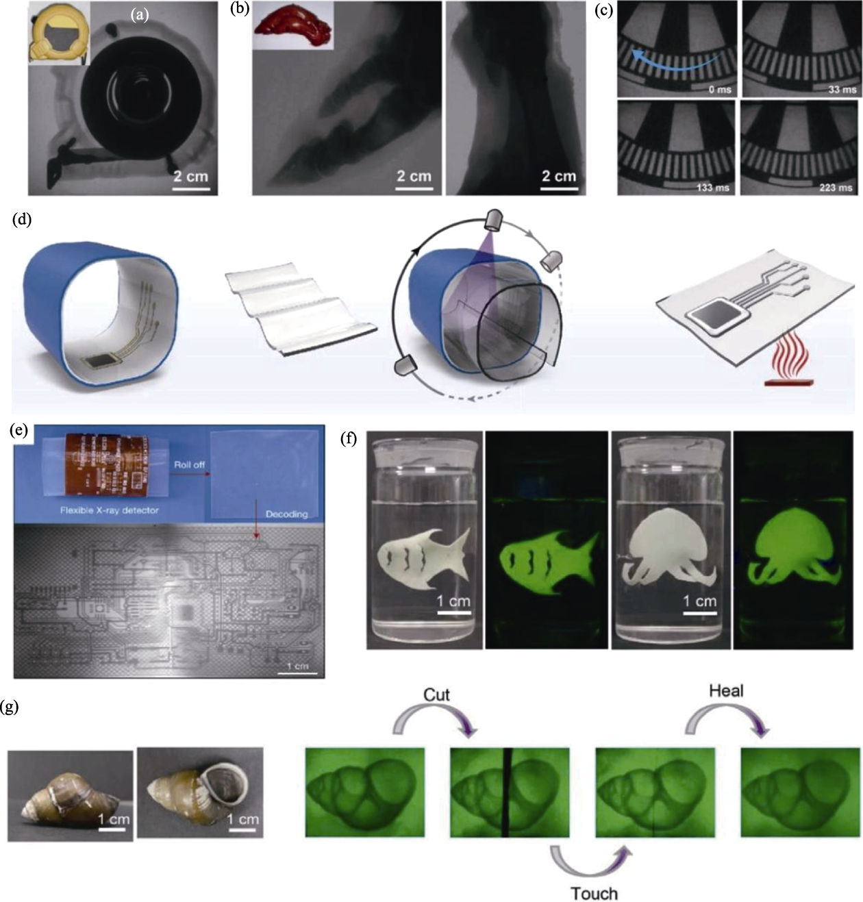
Fig. 6 XP materials for X-ray imaging applications[26,28,36] (a) X-ray image of a tape measure[28]; (b) Pig trotters with a large flashing screen[28]; (c) Dynamic X-ray image of a chopper[28]; (d) High resolution Xr LEI illustrated by schematic diagram of 3D electronic imaging achieved by a flexible detector integrated with nano scintillators (Firstly, insert the detector into the 3D electronic circuit board for conformal coating; Next, the image of the electronic board is projected onto the detector; After stopping the X-ray, the detector is transferred to a hot substrate for thermal stimulation and subsequent luminescence imaging)[36]; (e) Xr LEI (voltage 50 kV) of 3D electronic board using prototype NaLuF4:Tb (15%, in mol) @NaYF4 detector[36]; (f) Photos of organic gel scintillators immersed in water under visible light and 365 nm ultraviolet light[26]; (g) Photos of snails under visible light and organic gel scintillators conducting self-healing X-ray imaging in water[26]
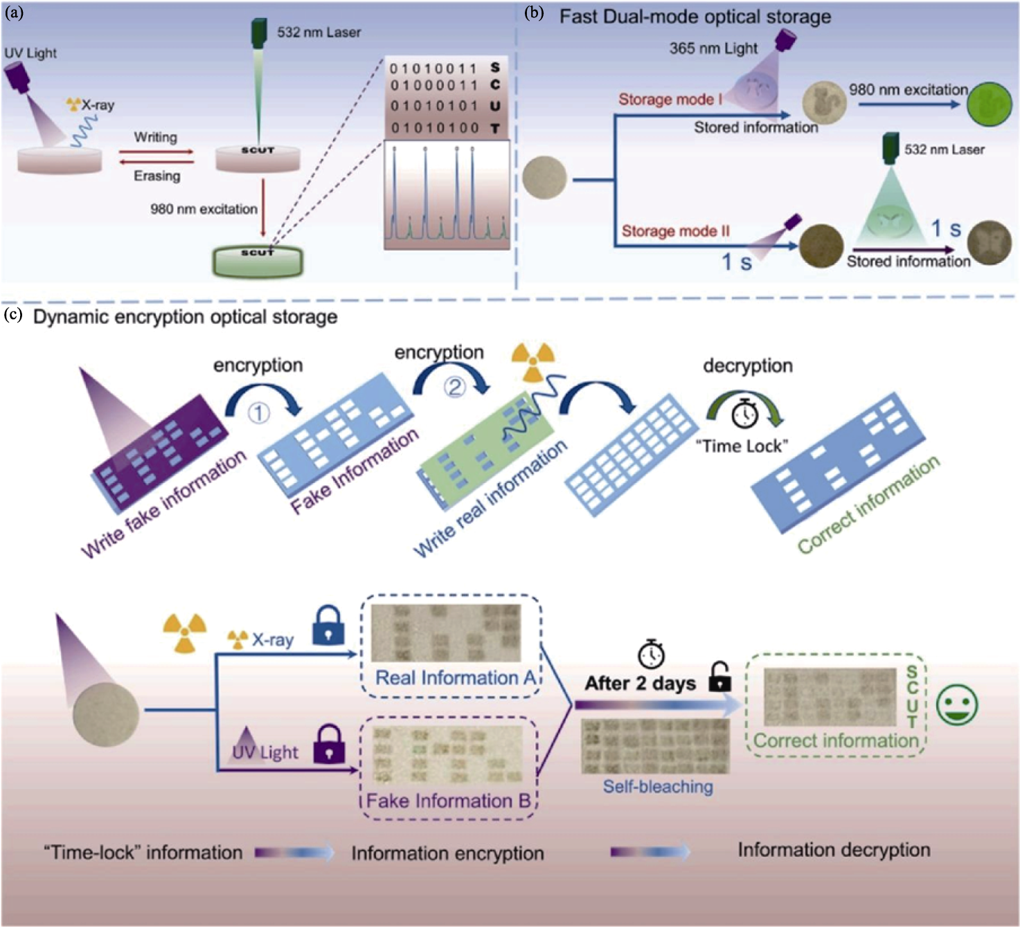
Fig. 7 Application of XP materials in the field of information storage and anti-counterfeiting[35] (a) BNN:Er ceramic for reading, writing, and erasing processes of optical information; (b) Dual mode for fast optical storage; (c) Dynamic encryption for optical storage model
| [1] |
GLASS B. The genetic hazards of nuclear radiations. Science, 1957, 126(3267): 241.
PMID |
| [2] | DU M H, WANG J, XU S J, et al. Super-elastic scintillating fibers and fabrics for efficient and visual radiation detection. Advanced Fiber Materials, 2023, 5(4): 1493. |
| [3] | LUO Z C, WU Y Y, WANG Y N, et al. Clinical radiation dose verification by topographic persistent luminescence dosimetry. Nano Today, 2023, 50: 101854. |
| [4] | CAO C T, TONEY M F, SHAM T K, et al. Emerging X-ray imaging technologies for energy materials. Materials Today, 2020, 34: 132. |
| [5] | HAN J S, LEE S H, GO H, et al. High-performance cold cathode X-ray tubes using a carbon nanotube field electron emitter. ACS Nano, 2022, 16(7): 10231. |
| [6] | NAKAJIMA T, MURAYAMA Y, MATSUZAWA T, et al. Development of a new highly sensitive LiF thermoluminescence dosimeter and its applications. Nuclear Instruments and Methods, 1978, 157(1): 155. |
| [7] | ZHANG H, YANG Z, ZHOU M, et al. Reproducible X-ray imaging with a perovskite nanocrystal scintillator embedded in a transparent amorphous network structure. Advanced Materials, 2021, 33(40): 2102529. |
| [8] | COLETTE D, MAZON D, BARNSLEY R, et al. Conceptual study of energy resolved X-ray measurement and electron temperature reconstruction on ITER with low voltage ionization chambers. The Review of Scientific Instruments, 2021, 92(8): 083511. |
| [9] | JIANG J, XIONG M, FAN K, et al. Synergistic strain engineering of perovskite single crystals for highly stable and sensitive X-ray detectors with low bias imaging and monitoring. Nature Photonics, 2022, 16: 575. |
| [10] |
THIRIMANNE H M, JAYAWARDENA K I, PARNELL A J, et al. High sensitivity organic inorganic hybrid X-ray detectors with direct transduction and broadband response. Nature Communications, 2018, 9: 2926.
DOI PMID |
| [11] | LEISTNER A L, PIANOWSKI Z L. Smart photochromic materials triggered with visible light. European Journal of Organic Chemistry, 2022, 2022(19): e202101271. |
| [12] | BEAUMONT J H, HART M. Multiple Bragg reflection monochromators for synchrotron X radiation. Journal of Physics E: Scientific Instruments, 1974, 7(10): 823. |
| [13] | KAYANI A B, KURIAKOSE S, MONSHIPOURI M, et al. UV photochromism in transition metal oxides and hybrid materials. Small, 2021, 17(32): 2100621. |
| [14] | WANG X, SHI H F, MA H L, et al. Organic phosphors with bright triplet excitons for efficient X-ray-excited luminescence. Nature Photonics, 2021, 15(3): 187. |
| [15] | KAWAMURA I, KAWAMOTO H, FUJIMOTO Y, et al. Isomerization behavior of diarylethene-type photochromic compounds under X-ray irradiation: application to dosimetry. Japanese Journal of Applied Physics, 2020, 59(4): 046004. |
| [16] | YANG D D, ZHENG H W, XUE J H, et al. Effect of lattice water on the photochromic property/photochromism of a Zn(II)-viologen coordination polymers. Journal of Molecular Structure, 2024, 1311: 138340. |
| [17] | WU J B, TAO C Y, LI Y, et al. Methylviologen-templated layered bimetal phosphate: a multifunctional X-ray-induced photochromic material. Chemical Science, 2014, 5(11): 4237. |
| [18] | HAN Y F, XU X M, WANG S H, et al. Reusable radiochromic semiconductive MOF for dual-mode X-ray detection using color change and electric signal. Chemical Engineering Journal, 2022, 437: 135468. |
| [19] |
LU L, PENG S C, ZHAO L, et al. Visualized X-ray dosimetry for multienvironment applications. Nano Letters, 2023, 23(18): 8753.
DOI PMID |
| [20] | YUAN M, JIA R, JIANG X, et al. Room-temperature X-ray induced photochromic properties of tungsten oxide. Materials Letters, 2023, 349: 134688. |
| [21] | YANG Z T, HU J Q, VAN DER HEGGEN D, et al. A versatile photochromic dosimeter enabling detection of X-Ray, ultraviolet, and visible photons. Laser & Photonics Reviews, 2023, 17(5): 2200809. |
| [22] | LIU J J, LU Y W, LI J, et al. UV and X-ray dual photochromic properties of three CPs based on a new viologen ligand. Dyes and Pigments, 2020, 177: 108276. |
| [23] | LIU J J, LU Y W, LU W B. Sun, UV and X-ray triple photochromic properties of three coordination polymers based on 1, 1′-bis(3-carboxylatobenzyl)-4,4′-bipyridinium ligand. CrystEngComm, 2020, 22(12): 2121. |
| [24] | ASAI K, KOSHIMIZU M, FUJIMOTO Y, et al. Isomerization behavior of spiropyran-based compounds upon X-ray irradiation. Radiation Measurements, 2017, 106: 166. |
| [25] | DU J R, YANG Z T, LIN H W, et al. Inorganic photochromic materials: recent advances, mechanism, and emerging applications. Responsive Materials, 2024, 2(2): e20240004. |
| [26] | LIU L L, HU H J, PAN W T, et al. Robust organogel scintillator for self-healing and ultra-flexible X-ray imaging. Advanced Materials, 2024, 36(13): 2311206. |
| [27] | GUO P Y, SUN C, ZHANG N N, et al. An inorganic-organic hybrid photochromic material with fast response to hard and soft X-rays at room temperature. Chemical Communications, 2018, 54(36): 4525. |
| [28] | ZHANG F, ZHOU Y C, CHEN Z P, et al. Large-area X-ray scintillator screen based on cesium hafnium chloride microcrystals films with high sensitivity and stability. Laser & Photonics Reviews, 2023, 17(5): 2200848. |
| [29] | SUN F, XU H, HONG W, et al. 2D CuInP2Se6 in high-sensitivity UV-VIS and X-ray detection. Advanced Functional Materials, 2024, 34(22): 2313776. |
| [30] | BYRON H C, SWAIN C, PATURI P, et al. Highly tuneable photochromic sodalites for dosimetry, security marking and imaging. Advanced Functional Materials, 2023, 33(42): 2303398. |
| [31] | SAMOILENKO Y, KAGANOVSKII Y, LIPOVSKII A A, et al. CW laser discoloration of X-ray irradiated silver doped silicate glasses. Optical Materials, 2008, 30(11): 1715. |
| [32] | YUAN W N, NIU G D, XIAN Y M, et al. In situ regulating the order-disorder phase transition in Cs2AgBiBr6 single crystal toward the application in an X-ray detector. Advanced Functional Materials, 2019, 29(20): 1900234. |
| [33] | TANG H T, LIU S B, FANG Z H, et al. High-resolution X-ray time-lapse imaging from fluoride nanocrystals embedded in glass matrix. Advanced Optical Materials, 2022, 10(12): 2102836. |
| [34] | SONG Y Y, ZHAO H P, ZI Y Z, et al. X-ray-irradiation-induced discoloration and persistent radioluminescence for reversible dual-mode imaging and detection applications. ACS Energy Letters, 2023, 8(5): 2232. |
| [35] | HU Y J, SUN Y S, HOU S R, et al. UV and X-ray induced photochromic material based on defect state exchanges. Chemical Engineering Journal, 2024, 495: 153600. |
| [36] | OU X Y, QIN X, HUANG B L, et al. High-resolution X-ray luminescence extension imaging. Nature, 2021, 590(7846): 410. |
| [37] | ZUO J, KEIL P, GRUNDMEIER G. Synthesis and characterization of photochromic Ag-embedded TiO2 nanocomposite thin films by non-reactive RF-magnetron sputter deposition. Applied Surface Science, 2012, 258(18): 7231. |
| [38] | HOLTON W C, BLUM H. Paramagnetic resonance of F centers in alkali halides. Physical Review, 1962, 125(1): 89. |
| [39] | SEITZ F. Color centers in alkali halide crystals. Reviews of Modern Physics, 1946, 18(3): 384. |
| [40] | JOSHI C, ABINANDANAN T A, MUKHERJEE R, et al. Destabilisation of nanoporous membranes through GB grooving and grain growth. Computational Materials Science, 2017, 139: 75. |
| [41] | ZHU Y, SUN H Q, JIA Q N, et al. Site-selective occupancy of Eu2+ toward high luminescence switching contrast in BaMgSiO4-based photochromic materials. Advanced Optical Materials, 2021, 9(6): 2001626. |
| [42] | JIN Y H, HU Y H, YUAN L F, et al. Multifunctional near-infrared emitting Cr3+-doped Mg4Ga8Ge2O20 particles with long persistent and photostimulated persistent luminescence, and photochromic properties. Journal of Materials Chemistry C, 2016, 4(27): 6614. |
| [43] | RABIN H, KLICK C C. Formation of F centers at low and room temperatures. Physical Review, 1960, 117(4): 1005. |
| [44] | ROY R. ChemInform abstract: ceramics by the solution-Sol-Gel route. ChemInform, 1988, 19(18): 1664. |
| [45] | BAI X, XU Z, ZI Y Z, et al. Dual-functional X-ray photochromic phosphor: high-performance detection and 3D imaging. Advanced Functional Materials, 2024, 34(37): 2402452. |
| [46] | PATHAK N, GUPTA S K, GHOSH P S, et al. Probing local site environments and distribution of manganese in SrZrO3:Mn; PL and EPR spectroscopy complimented by DFT calculations. RSC Advances, 2015, 5(23): 17501. |
| [47] | ZHANG Y Y, LUO L H, LI K X, et al. Reversible up-conversion luminescence modulation based on UV-VIS light-controlled photochromism in Er3+ doped Sr2SnO4+. Journal of Materials Chemistry C, 2018, 6(48): 13148. |
| [48] | LEE S H, CHEONG H M, LIU P, et al. Raman spectroscopic studies of gasochromic a-WO3 thin films. Electrochimica Acta, 2001, 46(13/14): 1995. |
| [49] | HE Y P, WU Z Y, FU L M, et al. Photochromism and size effect of WO3 and WO3-TiO2 aqueous sol. Chemistry of Materials, 2003, 15(21): 4039. |
| [50] | EGRANOV A V, SIZOVA T Y, SHENDRIK R Y, et al. Instability of some divalent rare earth ions and photochromic effect. Journal of Physics and Chemistry of Solids, 2016, 90: 7. |
| [51] | SCOULER W J, SMAKULA A. Coloration of pure and doped calcium fluoride crystals at 20 ℃ and -190 ℃. Physical Review, 1960, 120(4): 1154. |
| [52] | SUN Y H, LI C L, WANG W F, et al. A photochromic and scintillation Eu-MOF with visual X-ray detection in bright and dark environments. Chemical Communications, 2022, 58(25): 4056. |
| [53] | PAN L, LIU Z, WELTON C, et al. Ultrahigh-flux X-ray detection by a solution-grown perovskite CsPbBr3 single-crystal semiconductor detector. Advanced Materials, 2023, 35(25): 2211840. |
| [54] | YANG L L, ZHANG H, ZHOU M, et al. High-stable X-ray imaging from all-inorganic perovskite nanocrystals under a high dose radiation. The Journal of Physical Chemistry Letters, 2020, 11(21): 9203. |
| [55] | LIU J Y, SHABBIR B, WANG C J, et al. Flexible, printable soft-X-ray detectors based on all-inorganic perovskite quantum dots. Advanced Materials, 2019, 31(30): 1901644. |
| [56] |
ZHANG Y H, SUN R J, OU X Y, et al. Metal halide perovskite nanosheet for X-ray high-resolution scintillation imaging screens. ACS Nano, 2019, 13(2): 2520.
DOI PMID |
| [57] | LI L Q, TAO L T, WANG L X, et al. Monolithic integration of perovskite heterojunction on TFT backplanes through vapor deposition for sensitive and stable X-ray imaging. Science Advances, 2024, 10(17): eadj8659. |
| [58] | ZHAO J J, ZHAO L, DENG Y H, et al. Perovskite-filled membranes for flexible and large-area direct-conversion X-ray detector arrays. Nature Photonics, 2020, 14: 612. |
| [59] | LI W, XU Y L, PENG J L, et al. Evaporated perovskite thick junctions for X-ray detection. ACS Applied Materials & Interfaces, 2021, 13(2): 2971. |
| [60] | QIAN W, XU X W, WANG J, et al. An aerosol-liquid-solid process for the general synthesis of halide perovskite thick films for direct-conversion X-ray detectors. Matter, 2021, 4(3): 942. |
| [61] | LIU L L, LI W J, FENG X P, et al. Energy transfer assisted fast X-ray detection in direct/indirect hybrid perovskite wafer. Advanced Science, 2022, 9(15): 2103735. |
| [62] | XIA M L, SONG Z H, WU H D, et al. Compact and large-area perovskite films achieved via soft-pressing and multi-functional polymerizable binder for flat-panel X-ray imager. Advanced Functional Materials, 2022, 32(16): 2110729. |
| [63] | DEUMEL S, VAN BREEMEN A, GELINCK G, et al. High- sensitivity high-resolution X-ray imaging with soft-sintered metal halide perovskites. Nature Electronics, 2021, 4: 681. |
| [64] | DU X Y, LIU Y M, PAN W C, et al. Chemical potential diagram guided rational tuning of electrical properties: a case study of CsPbBr3 for X-ray detection. Advanced Materials, 2022, 34(17): 2110252. |
| [65] | ZHOU Y, ZHAO L, NI Z Y, et al. Heterojunction structures for reduced noise in large-area and sensitive perovskite X-ray detectors. Science Advances, 2021, 7(36): eabg6716. |
| [66] | CHEN C, SUN J K, ZHANG Y J, et al. Flexible viologen-based porous framework showing X-ray induced photochromism with single-crystal-to-single-crystal transformation. Angewandte International Edition Chemie, 2017, 56(46): 14458. |
| [67] |
LU H J, XIE J, WANG X Y, et al. Visible colorimetric dosimetry of UV and ionizing radiations by a dual-module photochromic nanocluster. Nature Communications, 2021, 12: 2798.
DOI PMID |
| [68] |
ABDOLLAHI A, ROGHANI-MAMAQANI H, RAZAVI B, et al. Photoluminescent and chromic nanomaterials for anticounterfeiting technologies: recent advances and future challenges. ACS Nano, 2020, 14(11): 14417.
DOI PMID |
| [69] | BAR N, CHOWDHURY P. A brief review on advances in rhodamine B based chromic materials and their prospects. ACS Applied Electronic Materials, 2022, 4(8): 3749. |
| [70] |
WUTTIG M, YAMADA N. Phase-change materials for rewriteable data storage. Nature Materials, 2007, 6(11): 824.
PMID |
| [71] | SUN H Q, LI X F, ZHU Y, et al. Achieving multicolor emission readout and tunable photoswitching via multiplexing of dual lanthanides in ferroelectric oxides. Journal of Materials Chemistry C, 2019, 7(19): 5782. |
| [72] | YANG Z T, DU J R, MARTIN L I D J, et al. Designing photochromic materials with large luminescence modulation and strong photochromic efficiency for dual-mode rewritable optical storage. Advanced Optical Materials, 2021, 9(20): 2100669. |
| [73] | LI X F, GUAN L L, LI Y, et al. Optical control of Er3+-doped M0.5Bi2.5Nb2O9 (M=Li, Na, K) materials for thermal stability and temperature sensing using photochromic reactions. Journal of Materials Chemistry C, 2020, 8(44): 15685. |
| [74] | ZHANG H L, ZHANG X, SUN W H, et al. All-solid-state transparent variable infrared emissivity devices for multi-mode smart windows. Advanced Functional Materials, 2024, 34(16): 2307356. |
| [75] | CHOI K, CHON J W M, GU M, et al. Low-distortion holographic data storage media using free-radical ring-opening polymerization. Advanced Functional Materials, 2009, 19(22): 3560. |
| [76] | XIAO Y, XIONG P, ZHANG S, et al. Cation-defect-induced self-reduction towards efficient mechanoluminescence in Mn2+- activated perovskites. Materials Horizons, 2023, 10(9): 3476. |
| [1] | LIU Leimin, LUO Hongxin, HE Yumei, JIN Limin, LI Yongjie, LIU Jingwen, WEI Yuquan, SUN Anle, CHEN Zhongming, LIU Xuejian, YIN Jie, HUANG Zhengren. Performance of Silicon Carbide Mirrors for Advanced Light Source Devices [J]. Journal of Inorganic Materials, 2026, 41(6): 805-813. |
| [2] | CHEN Mingjun, MIAO Hongkang, XIAO Yingjun, DENG Jianbo, ZHANG Xiang, ZHAO Jiupeng, LI Yao. Photo- and Thermo-chromic Dual-responsive Materials: A Review on Design Strategies and Applications in Smart Windows [J]. Journal of Inorganic Materials, 2026, 41(6): 723-738. |
| [3] | SONG Kunjie, XIE Rongjun. Research Advances on Machine Learning-driven Development of Novel Luminescent Materials [J]. Journal of Inorganic Materials, 2026, 41(6): 689-703. |
| [4] | HU Yuqing, ZHU Yixin, LE Xianhao, WAN Qing. Lithium Tantalate Wafer: Advances in Thinning Technology and Application in Pyroelectric Infrared Detectors [J]. Journal of Inorganic Materials, 2026, 41(6): 764-774. |
| [5] | LIU Chunfan, CHEN Ke, GE Fangfang, HUANG Qing. Research Progress on Lead-bismuth Eutectic Corrosion Resistant Coatings [J]. Journal of Inorganic Materials, 2026, 41(6): 775-786. |
| [6] | HU Yang, XIE Min, ZHANG Xiaoyi, LI Xiang, GUO Xinwei, JIANG Nan, ZHOU Wenhan, ZHANG Shengli, ZENG Haibo. Research Progress on Computational and Data-driven Environmental-friendly Luminescent Materials [J]. Journal of Inorganic Materials, 2026, 41(6): 704-722. |
| [7] | WANG Junbu, HUANG Zeai, YANG Mingkai, MENG Ying, ZHOU Mingwei, ZHOU Ying. Research Progress on Anti-coking Catalytic Materials for Methane Conversion [J]. Journal of Inorganic Materials, 2026, 41(6): 739-750. |
| [8] | WANG Jinwen, YANG Zhen, ZHOU Huan, XIA Dan, YANG Lei. Biomedical Applications of Injectable Inorganic Biomaterials [J]. Journal of Inorganic Materials, 2026, 41(6): 751-763. |
| [9] | LI Hantao, SHEN Qiang, LUO Guoqiang, WANG Xuefei, GAO Ming, CHEN Chen. Research Progress on Structure and Performance Regulation of Silicon-based Anode Materials via Mechanical Ball Milling [J]. Journal of Inorganic Materials, 2026, 41(5): 561-572. |
| [10] | XIE Chenyi, MIAO Huaming, ZHANG Weiran, LIU Rongjun, WANG Yanfei, LI Duan. Research Progress on Theoretical Calculation in the Field of High-entropy Ceramics [J]. Journal of Inorganic Materials, 2026, 41(5): 545-560. |
| [11] | LI Xuan, YE Kuicai, FENG Jiayin, QIU Jiajun, QIAN Wenhao, XING Min. Surface Modification of Titanium-based Dental Implants for Soft Tissue Sealing: A Review [J]. Journal of Inorganic Materials, 2026, 41(4): 432-444. |
| [12] | PENG Dezhao, LI Rui, WANG Wenhong, WANG Zirui, ZHANG Zhizhen. Research Progress on Sodium Chloride Solid Electrolytes [J]. Journal of Inorganic Materials, 2026, 41(4): 409-420. |
| [13] | CHEN Kun, JIANG Yonggang, FENG Junzong, LI Liangjun, HU Yijie, FENG Jian. Research Progress on Lanthanum Zirconate Porous Materials for Thermal Insulation [J]. Journal of Inorganic Materials, 2026, 41(4): 421-431. |
| [14] | WEI Lianjin, QI Zhijie, WANG Xin, ZHU Junwu, FU Yongsheng. Modification of Nanodiamond and Its Application in Electrocatalytic Oxygen Reduction Reaction [J]. Journal of Inorganic Materials, 2026, 41(3): 273-288. |
| [15] | LIU Zhanyi, LI Mian, OUYANG Xiaoping, CHAI Zhifang, HUANG Qing. Recent Progress on Removal of Sr/Cs from Molten Salt in Dry Reprocessing [J]. Journal of Inorganic Materials, 2026, 41(2): 150-158. |
| Viewed | ||||||
|
Full text |
|
|||||
|
Abstract |
|
|||||