Journal of Inorganic Materials ›› 2023, Vol. 38 ›› Issue (1): 3-31.DOI: 10.15541/jim20220218
Special Issue: 【信息功能】敏感陶瓷(202512)
• Topical Section: Anti-epidemic Biomaterials (Contributing Editor: YANG Yong) • Previous Articles Next Articles
LI Yanyan1,2( ), PENG Yusi1,2, LIN Chenglong1,2, LUO Xiaoying3, TENG Zheng4(), ZHANG Xi4, HUANG Zhengren1,2, YANG Yong1,2()
Received:2022-04-12
Revised:2022-05-03
Published:2023-01-20
Online:2022-06-22
Contact:
YANG Yong, professor. E-mail: yangyong@mail.sic.ac.cn;About author:LI Yanyan (1997-), female, PhD candidate. E-mail: liyanyan20@mails.ucas.ac.cn
Supported by:CLC Number:
LI Yanyan, PENG Yusi, LIN Chenglong, LUO Xiaoying, TENG Zheng, ZHANG Xi, HUANG Zhengren, YANG Yong. Nanomaterials and Biosensing Technology for the SARS-CoV-2 Detection[J]. Journal of Inorganic Materials, 2023, 38(1): 3-31.
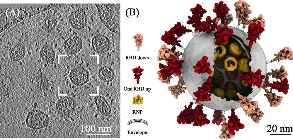
Fig. 1 TEM image (A) and schemetic morphology (B) of SARS-CoV-2[9] The color figures can be obtained from online edition
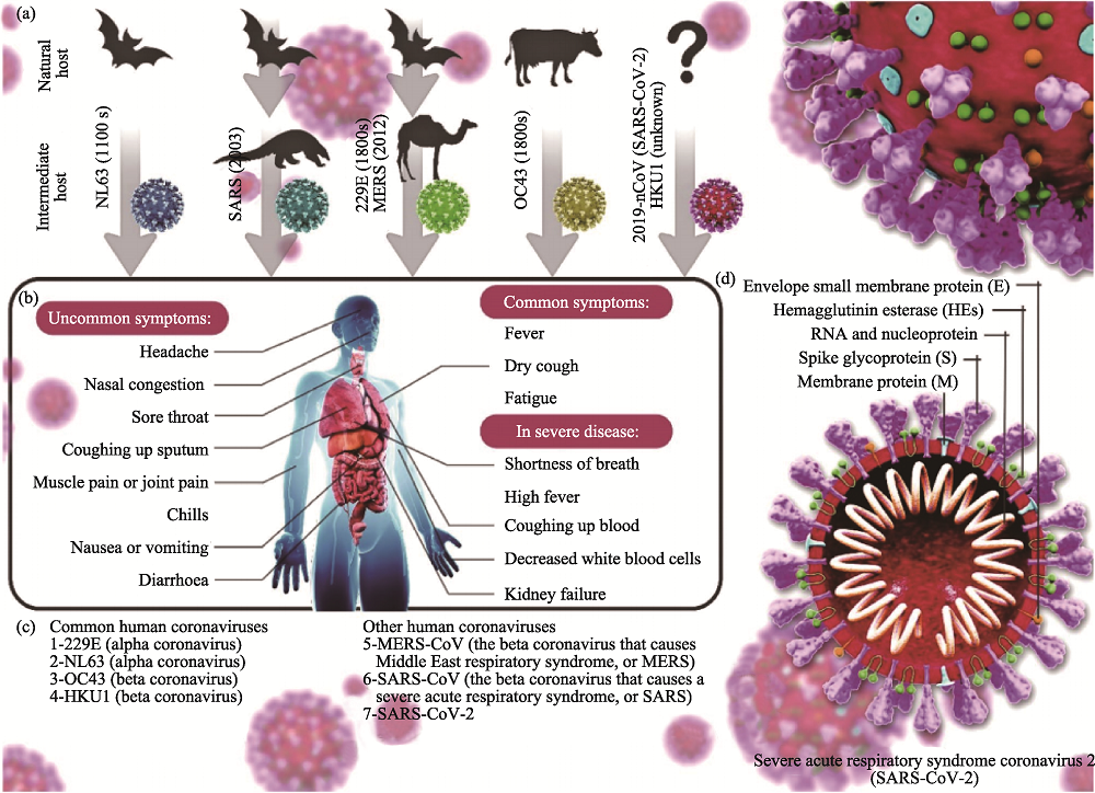
Fig. 2 Classification (a, c), symptoms (b) and structure (c) of coronavirus[16] The color figures can be obtained from online edition
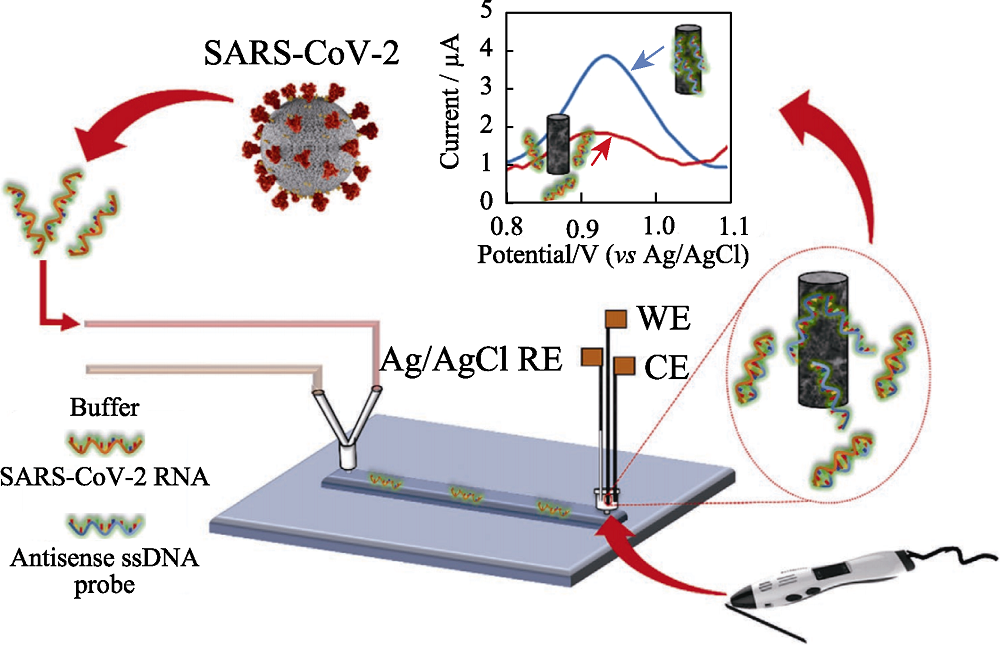
Fig. 3 Scheme of the lab-on-a-chip genosensor for SARS- CoV-2 virus detection[35] WE: working electrode; CE: counter electrode; RE: reference electrode; ssDNA: single strand DNA The color figure can be obtained from online edition
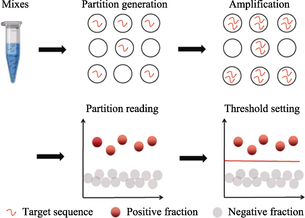
Fig. 4 Principle workflow of ddPCR in analyzing clinical samples[37] The color figure can be obtained from online edition
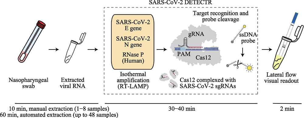
Fig. 5 Detection of SARS-CoV-2 based on CRISPR-Cas12 combined with lateral flow technique[44] The color figure can be obtained from online edition
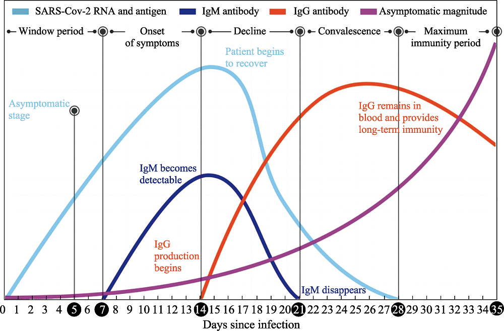
Fig. 6 Change of human immune response after viral infection[16] The color figure can be obtained from online edition
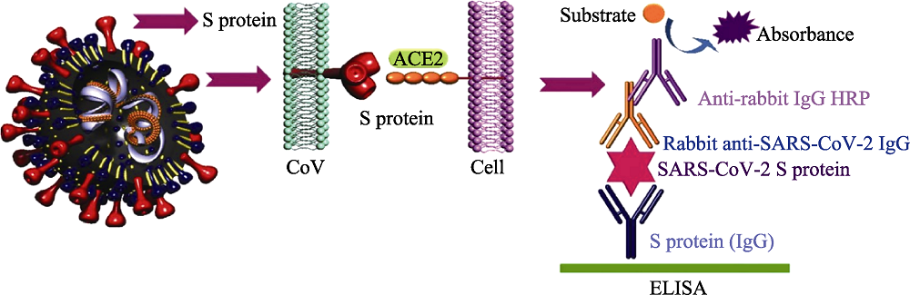
Fig. 7 ELISA method for detection of SARS-CoV-2[16] ACE2: angiotensin converting enzyme 2; HRP: horse radish peroxidase; ELISA: enzyme linked immunosorbent assay The color figure can be obtained from online edition
| Object sample | Characteristic | Detection technology | Advantage | Disadvantage |
|---|---|---|---|---|
| RNA | 1. Target: the gene sequences of SARS-CoV-2 2. Great producibility 3. Long detection period 4. Possibility of being contaminated and false positive result | Whole genome sequencing | 1. High accuracy and sensitivity 2. Reflecting genetic information of pathogen comprehensively | 1. Expensive special instruments 2. Relying on professionals 3. Difficulty in detection on a large scale |
| RT-qPCR | 1. High sensitivity and specificity 2. Low cost | 1. Long amplification time 2. High requirements of equipment 3. Complex operation | ||
| LAMP | 1. Isothermal reaction 2. High efficiency and speed 3. High sensitivity and visualization | 1. Complex design of primer 2. Low specificity | ||
| Microfluidic chip | 1. Multiple detection of pathogens 2. Integration of sample preparation and detection 3. Ability in automate analysis | Difficulty in chip design, material selection, processing, packaging, and storage | ||
| ddPCR | 1. High sensitivity and lowest limitation of detection 2. Facilitation and high degree of automation 3. Quantitative detection | 1. Small reaction volume 2. Expensive equipment and reagents | ||
| CRISPR | 1. High speed and low cost 2. High sensitivity 3. Strong system stability 4. On-site detection | The accuracy of detection needs to be verified | ||
| Antibodies | 1. Target: human antibodies stimulated by SARS-CoV-2 2. Easy sample collection and low detection threshold 3. Simple operation and high throughput 4. Limitation of timeframe 5. Lower sensitivity and specificity than those of nucleic acid detection | ELISA | 1. Low difficulty of standardization of carrier 2. High sensitivity and specificity 3. Simple equipment | 1. Long detection time and cumbersome steps 2. Limited single detection throughout |
| LFIA (Colloidal gold method) | 1. On-site detection caused by easy operation 2. High sensitivity and speed 3. Low cost 4. Mass production | 1. Only qualitative analysis 2. Different reproducibility of different batches of products | ||
| CLIA | 1. High sensitivity and specificity 2. High throughput detection and high degree of automation | 1. Special instrument 2. High detection cost | ||
| Antigen | 1. Target: SARS-CoV-2 antigen 2. Simple and fast operation | LFIA (Colloidal gold method) | 1. Fast and facile operation 2. Visualization 3. On-site detection and large-scale population screening | Low sensitivity |
Table 1 Comparison of conventional detection methods for SARS-CoV-2
| Object sample | Characteristic | Detection technology | Advantage | Disadvantage |
|---|---|---|---|---|
| RNA | 1. Target: the gene sequences of SARS-CoV-2 2. Great producibility 3. Long detection period 4. Possibility of being contaminated and false positive result | Whole genome sequencing | 1. High accuracy and sensitivity 2. Reflecting genetic information of pathogen comprehensively | 1. Expensive special instruments 2. Relying on professionals 3. Difficulty in detection on a large scale |
| RT-qPCR | 1. High sensitivity and specificity 2. Low cost | 1. Long amplification time 2. High requirements of equipment 3. Complex operation | ||
| LAMP | 1. Isothermal reaction 2. High efficiency and speed 3. High sensitivity and visualization | 1. Complex design of primer 2. Low specificity | ||
| Microfluidic chip | 1. Multiple detection of pathogens 2. Integration of sample preparation and detection 3. Ability in automate analysis | Difficulty in chip design, material selection, processing, packaging, and storage | ||
| ddPCR | 1. High sensitivity and lowest limitation of detection 2. Facilitation and high degree of automation 3. Quantitative detection | 1. Small reaction volume 2. Expensive equipment and reagents | ||
| CRISPR | 1. High speed and low cost 2. High sensitivity 3. Strong system stability 4. On-site detection | The accuracy of detection needs to be verified | ||
| Antibodies | 1. Target: human antibodies stimulated by SARS-CoV-2 2. Easy sample collection and low detection threshold 3. Simple operation and high throughput 4. Limitation of timeframe 5. Lower sensitivity and specificity than those of nucleic acid detection | ELISA | 1. Low difficulty of standardization of carrier 2. High sensitivity and specificity 3. Simple equipment | 1. Long detection time and cumbersome steps 2. Limited single detection throughout |
| LFIA (Colloidal gold method) | 1. On-site detection caused by easy operation 2. High sensitivity and speed 3. Low cost 4. Mass production | 1. Only qualitative analysis 2. Different reproducibility of different batches of products | ||
| CLIA | 1. High sensitivity and specificity 2. High throughput detection and high degree of automation | 1. Special instrument 2. High detection cost | ||
| Antigen | 1. Target: SARS-CoV-2 antigen 2. Simple and fast operation | LFIA (Colloidal gold method) | 1. Fast and facile operation 2. Visualization 3. On-site detection and large-scale population screening | Low sensitivity |
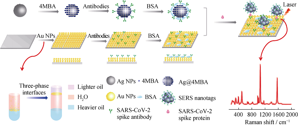
Fig. 8 Schematic illustration of the SERS-based immunoassay[77] MBA: thiosalicylic acid; BSA: bovine serum albumin; NPs: nanoparticles The color figure can be obtained from online edition
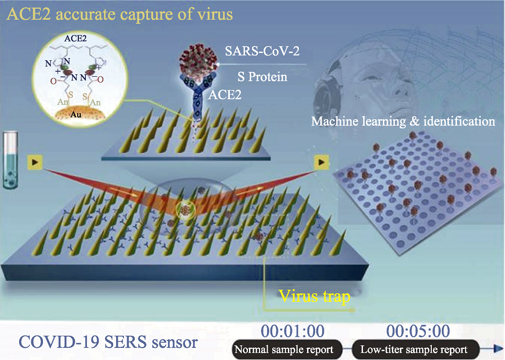
Fig. 9 Schematic diagram of operation procedure of COVID-19 SERS sensor[8] The color figure can be obtained from online edition
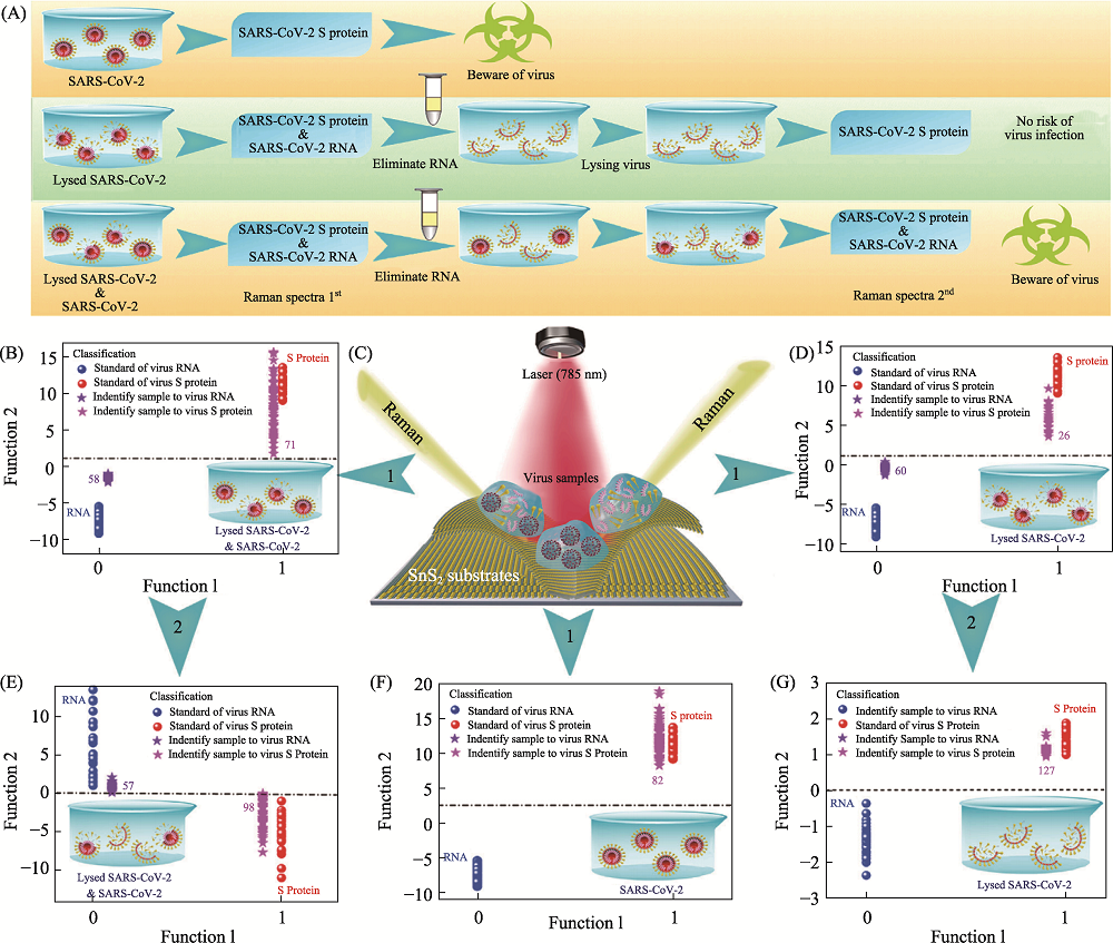
Fig. 10 Application of SnS2 microspheres for diagnosing the infectiousness of SARS-CoV-2[80] (A) Experimental procedure for diagnosing the infectiousness of SARS-CoV-2; (B) SVM analysis results to identify the mixture of the SARS-CoV-2 with complete viral structure and the lysed SARS-CoV-2; (C) Raman scattering diagram of three contamination situations of the novel coronavirus based on SnS2 substrates; (D) SVM analysis results to identify the lysed SARS-CoV-2; (E) SVM analysis results to identify the mixture of the SARS-CoV-2 with complete viral structure and the lysed SARS-CoV-2 after eliminating RNA and relysing virus samples; (F) SVM analysis results to identify the SARS-CoV-2 with complete viral structure; (G) SVM analysis results to identify the lysed SARS-CoV-2 after eliminating RNA and relysing virus samples. SVM: support vector machine The color figure can be obtained from online edition
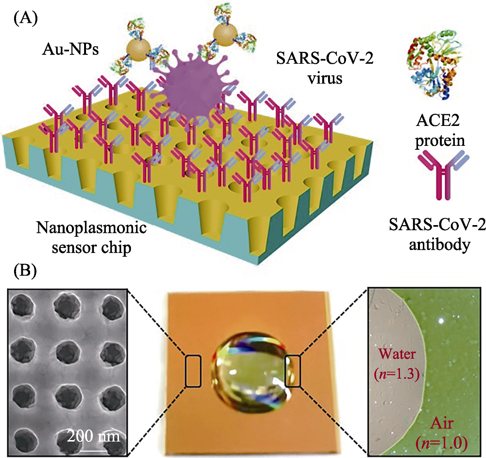
Fig. 11 Schematic diagram of nano-plasma optic sensor for detection of SARS-CoV-2[89] (A) Schematic diagram of the nanoplasmonic resonance sensor for determination of SARS-CoV-2 pseudovirus concentration; (B) Photograph (middle) of one piece of Au nanocup array chip with a drop of water on top The color figure can be obtained from online edition
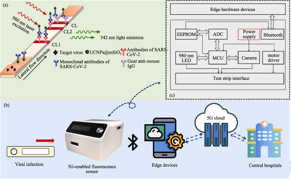
Fig. 12 SARS-CoV-2 detection based on 5G-enabled fluorescence biosensor[100] (a) The principle of the UCNPs based lateral flow assay in detection of SARS-CoV-2; (b) The working process of the proposed 5G-enabled fluorescence sensor; (c) The circuit configuration and hardware composition of the fluorescence sensor; CL: control line; TL1: test line 1; TL2: test line 2; UCNPs: up-conversion nanoparticles; EEPROM: electrically erasable programmable read only memory; ADC: application data center; MCU: motor control unit The color figure can be obtained from online edition
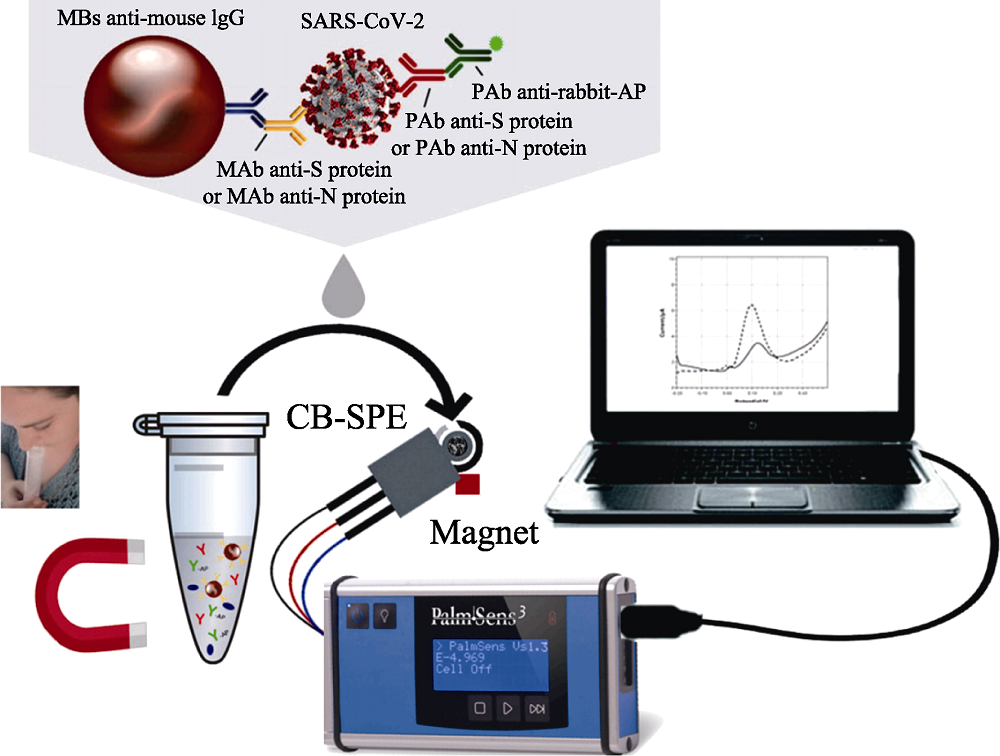
Fig. 13 Magnetic beads-based electrochemical assay for SARS-CoV-2 detection in untreated saliva[126] MBs: magnetic beads; MAb: monoclonal antibody; PAb: polyclonal antibody; AP: alkaline phosphatase; CB-SPE: carbon-based screen-printed electrodes The color figure can be obtained from online edition
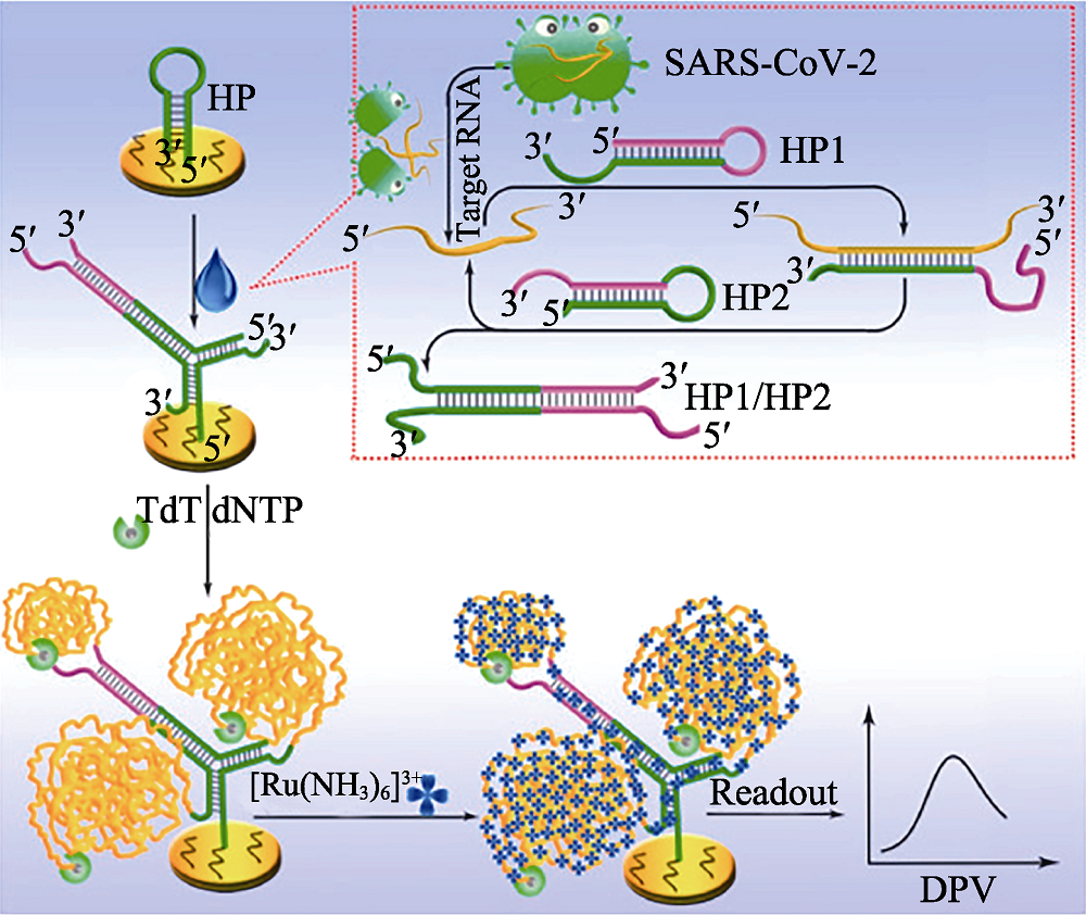
Fig. 14 Principle of the proposed electrochemical biosensor for sensitive analysis of SARS-CoV-2 RNA[136] HP: hairpin; TdT: terminal deoxynucleotidyl transferase; dNTP: deoxyribonucleotides; DPV: differential pulse voltammetry The color figure can be obtained from online edition
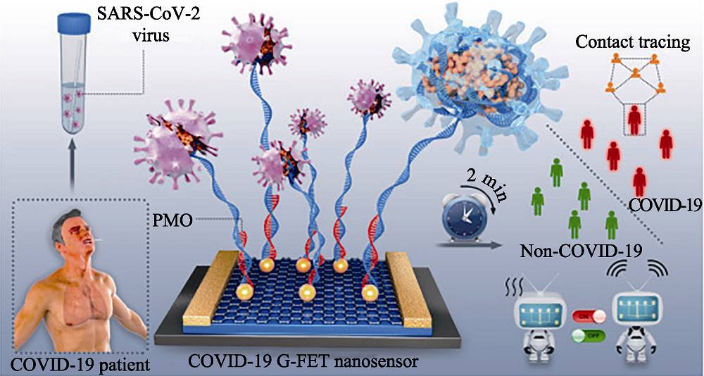
Fig. 15 Schematic diagram of rapid direct identification of SARS-CoV-2 using PMO-functionalized G-FET nano-sensors[148] G-FET: graphene field-effect transistor; PMO: phosphorodiamidate morpholino oligos The color figure can be obtained from online edition
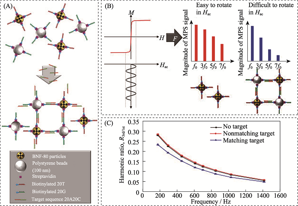
Fig. 16 Schematic diagram of detection of SARS-CoV-2 RNA based on magnetic particle spectroscopy biosensors[166] (A) Magnetic nanoparticles (gold) and polystyrene beads (silver) with streptavidin (purple)-modified surface are equipped with single stranded DNA strands (red and green, respectively) with a specific sequence via biotin-streptavidin-binding; (B) Applying a sinusoidal magnetic field (black) to a solution of nanoparticles results in reorientation of the nanoparticles which can be readout by measuring the magnetic response M of the nanoparticles; (C) Exemplary spectrum of the ratio of received harmonics as a function of excitation frequencies for 80 nm BNF magnetic particles The color figure can be obtained from online edition
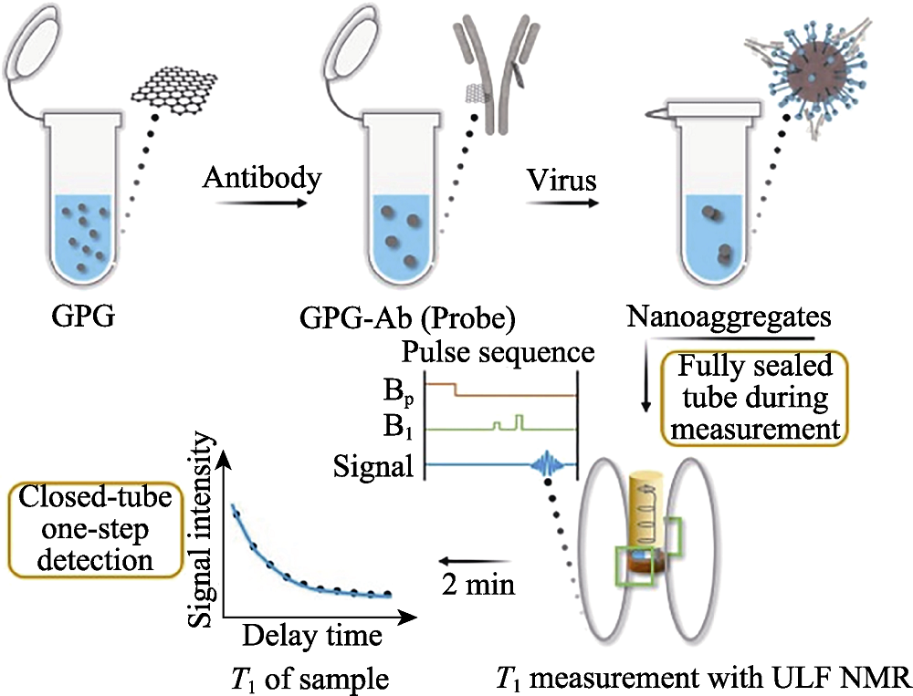
Fig. 17 Detection process of SARS-CoV-2 of the magnetic relaxation switches assay with ULF NMR[169] ULF: ultra-low field; GPG: Gd3+ loaded PEG modified GQDs; GQDs: graphene quantum dots The color figure can be obtained from online edition
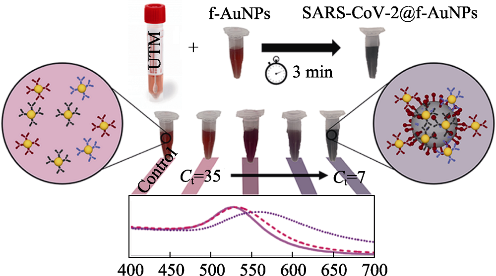
Fig. 18 Schematic diagram of the detection of SARS-CoV-2 based on colorimetric biosensors[187] UTM: universal transport medium The color figure can be obtained from online edition
| Detection technology | Detection method | Object | Sample | Related material | Detection time | Lower detection limit | Ref. |
|---|---|---|---|---|---|---|---|
| SERS-based biosensors | Labelled-SERS | S protein | Lysis solution | Macro/nanostructure Au substrate | 15 min | 10 PFU/mL | [ |
| Label-free SERS | Virus particles | Nasal/throat solution | Macro/nanostructure Au substrate, Au nanoparticles | 15 min | 60 copies/mL | [ | |
| SPR-based biosensors | Combining SPR and LSPR | Pseudovirus particles | N/A | Macro/nanostructure Au substrate, Au nanoparticles | 15 min | 370 vp/mL | [ |
| Fluorescence biosensors | “signal on” mode | RNA | Lysis solution | N/A | 15 samples/ 45 min | 600 copies/mL | [ |
| Electrochemical biosensors | Voltammetric/ amperometric biosensors | RNA | Nasal/throat solution | Au nanoparticles | 5 min | 6900 copies/mL | [ |
| Impedimetric biosensors | Antibodies | Serum | Au nanoparticles | 30 min | N/A | [ | |
| Potentiometric biosensors | Cholinesterase | Blood | Graphene and copper | ~7 s (only detection time) | 7.9 × 10⁻⁸ mol/L | [ | |
| FET-based biosensors | RNA | Nasal/throat solution | Graphene | 1 min (only detection time) | 10-20 copies/mL | [ | |
| Magnetic biosensors | Magnetoresistance | Antibodies | Blood | Magnetic nanoparticles | 10 min | 5-10 ng/mL | [ |
| Magnetic particle spectroscopy platforms | S protein and N protein | PBS | Magnetic nanoparticles | N/A | 1.56 nmol/L | [ | |
| Nuclear magnetic resonance | Antibodies | Blood | Magnetic graphene quantum dot | 2 min | 248 vp/mL | [ | |
| Colorimetric biosensors | Agglomeration of nanoparticles | RNA | N/A | Au nanoparticles | >45 min | 160 fmol/L | [ |
Table 2 Comparison of novel biosensors for SARS-CoV-2 detection
| Detection technology | Detection method | Object | Sample | Related material | Detection time | Lower detection limit | Ref. |
|---|---|---|---|---|---|---|---|
| SERS-based biosensors | Labelled-SERS | S protein | Lysis solution | Macro/nanostructure Au substrate | 15 min | 10 PFU/mL | [ |
| Label-free SERS | Virus particles | Nasal/throat solution | Macro/nanostructure Au substrate, Au nanoparticles | 15 min | 60 copies/mL | [ | |
| SPR-based biosensors | Combining SPR and LSPR | Pseudovirus particles | N/A | Macro/nanostructure Au substrate, Au nanoparticles | 15 min | 370 vp/mL | [ |
| Fluorescence biosensors | “signal on” mode | RNA | Lysis solution | N/A | 15 samples/ 45 min | 600 copies/mL | [ |
| Electrochemical biosensors | Voltammetric/ amperometric biosensors | RNA | Nasal/throat solution | Au nanoparticles | 5 min | 6900 copies/mL | [ |
| Impedimetric biosensors | Antibodies | Serum | Au nanoparticles | 30 min | N/A | [ | |
| Potentiometric biosensors | Cholinesterase | Blood | Graphene and copper | ~7 s (only detection time) | 7.9 × 10⁻⁸ mol/L | [ | |
| FET-based biosensors | RNA | Nasal/throat solution | Graphene | 1 min (only detection time) | 10-20 copies/mL | [ | |
| Magnetic biosensors | Magnetoresistance | Antibodies | Blood | Magnetic nanoparticles | 10 min | 5-10 ng/mL | [ |
| Magnetic particle spectroscopy platforms | S protein and N protein | PBS | Magnetic nanoparticles | N/A | 1.56 nmol/L | [ | |
| Nuclear magnetic resonance | Antibodies | Blood | Magnetic graphene quantum dot | 2 min | 248 vp/mL | [ | |
| Colorimetric biosensors | Agglomeration of nanoparticles | RNA | N/A | Au nanoparticles | >45 min | 160 fmol/L | [ |
| [1] |
YUCE M, FILIZTEKIN E, OZKAYA K G. COVID-19 diagnosis-a review of current methods. Biosensors and Bioelectronics, 2021, 172: 112752.
DOI URL |
| [2] |
JI T X, LIU Z W, WANG G Q, et al. Detection of COVID-19: a review of the current literature and future perspectives. Biosensors and Bioelectronics, 2020, 166: 112455.
DOI URL |
| [3] |
XUE X, BALL J K, ALEXANDER C, et al. All surfaces are not equal in contact transmission of SARS-CoV-2. Matter, 2020, 3(5): 1433.
DOI PMID |
| [4] |
CHEN W, CAI B, GENG Z, et al. Reducing false negatives in COVID-19 testing by using microneedle-based oropharyngeal swabs. Matter, 2020, 3(5): 1589.
DOI PMID |
| [5] |
CUI F Y, ZHOU H S. Diagnostic methods and potential portable biosensors for coronavirus disease 2019. Biosensors and Bioelectronics, 2020, 165: 112349.
DOI URL |
| [6] | LIN D C, LIU L, ZHANG M X, et al. Evaluations of the serological test in the diagnosis of 2019 novel coronavirus (SARS-CoV-2) infections during the COVID-19 outbreak. European Journal of Clinical Microbiology & Infectious Diseases, 2020, 39(12): 2271. |
| [7] |
SEO G, LEE G, KIM M J, et al. Correction to rapid detection of COVID-19 causative virus (SARS-CoV-2) in human nasopharyngeal swab specimens using field-effect transistor-based biosensor. ACS Nano, 2020, 14(9): 12257.
DOI PMID |
| [8] |
YANG Y, PENG Y S, LIN C L, et al. Human ACE2-functionalized gold “virus-trap” nanostructures for accurate capture of SARS-CoV-2 and single-virus SERS detection. Nano-Micro Letters, 2021, 13: 109.
DOI URL |
| [9] |
YAO H P, SONG Y T, CHEN Y, et al. Molecular architecture of the SARS-CoV-2 virus. Cell, 2020, 183(3): 730.
DOI PMID |
| [10] |
ZHOU P, YANG X L, WANG X G, et al. A pneumonia outbreak associated with a new coronavirus of probable bat origin. Nature, 2020, 579(7798): 270.
DOI URL |
| [11] |
LI W H, MOORE J M, VASILIEVA N, et al. Angiotensin-converting enzyme 2 is a functional receptor for the SARS coronavirus. Nature, 2003, 426: 450.
DOI URL |
| [12] |
HOFMANN H, PYRC K, HOEK V D L, et al. Human coronavirus NL63 employs the severe acute respiratory syndrome coronavirus receptor for cellular entry. Proceedings of the National Academy of Sciences of the United States of America, 2005, 102(22): 7988.
DOI PMID |
| [13] |
LU G W, HU Y W, WANG Q H, et al. Molecular basis of binding between novel human coronavirus MERS-CoV and its receptor CD26. Nature, 2013, 500(7461): 227.
DOI URL |
| [14] |
RAJ V S, MOU H, SMITS S L, et al. Dipeptidyl peptidase 4 is a functional receptor for the emerging human coronavirus-EMC. Nature, 2013, 495 (7440): 251.
DOI URL |
| [15] |
MENACHERY V D, YOUNT B L JR, DEBBINK K, et al. A SARS-like cluster of circulating bat coronaviruses shows potential for human emergence. Nature Medicine, 2015, 21(12): 1508.
DOI PMID |
| [16] |
OROOJI Y, SOHRABI H, HEMMAT N, et al. An overview on SARS-CoV-2 (COVID-19) and other human coronaviruses and their detection capability via amplification assay, chemical sensing, biosensing, immunosensing, and clinical assays. Nano-Micro Letters, 2021, 13: 18.
DOI URL |
| [17] |
TO K K W, TSANG O T Y, LEUNG W S, et al. Temporal profiles of viral load in posterior oropharyngeal saliva samples and serum antibody responses during infection by SARS-CoV-2: an observational cohort study. The Lancet Infectious Diseases, 2020, 20(5): 565.
DOI URL |
| [18] |
DONG X, CAO Y Y, LU X X, et al. Eleven faces of coronavirus disease 2019. Allergy, 2020, 75(7): 1699.
DOI PMID |
| [19] | CORMAN V M, LANDT O, KAISER M, et al. Detection of 2019 novel coronavirus (2019-nCoV) by real-time RT-PCR. Euro surveillnace, 2020, 25(3): 23. |
| [20] |
LU R J, ZHAO X, LI J, et al. Genomic characterisation and epidemiology of 2019 novel coronavirus: implications for virus origins and receptor binding. The Lancet, 2020, 395(10224): 565.
DOI URL |
| [21] |
REN L L, WANG Y M, WU Z Q, et al. Identification of a novel coronavirus causing severe pneumonia in human: a descriptive study. Chinese Medical Journal, 2020, 133(9): 1015.
DOI URL |
| [22] |
PEIRIS S M J, PHIL D, YUEN Y K, et al. The severe acute respiratory syndrome. The New England Journal of Medicine, 2003, 349: 2431.
DOI PMID |
| [23] | CHEN L J, LIU W Y, ZHANG Q, et al. RNA based mNGS approach identifies a novel human coronavirus from two individual pneumonia cases in 2019 Wuhan outbreak. Emerging Microbes & Infections, 2020, 9(1): 313. |
| [24] |
SHI M, ZHANG Y Z, HOLMES E C. Meta-transcriptomics and the evolutionary biology of RNA viruses. Virus Research, 2018, 243: 83.
DOI PMID |
| [25] |
SAKAMOTO Y, SEREEWATTANAWOOT S, SUZUKI A. A new era of long-read sequencing for cancer genomics. Journal of Human Genetics, 2020, 65(1): 3.
DOI PMID |
| [26] |
WANG M, FU A S, HU B, et al. Nanopore targeted sequencing for the accurate and comprehensive detection of SARS-CoV-2 and other respiratory viruses. Small, 2020, 16: 2002169.
DOI URL |
| [27] |
NOTOMI T, OKAYAMA H, MASUBUCHI H, et al. Loop-mediated isothermal amplification of DNA. Nucleic Acids Research, 2000, 28: 63.
PMID |
| [28] |
ZHU X, WANG X X, HAN L M, et al. Multiplex reverse transcription loop-mediated isothermal amplification combined with nanoparticle-based lateral flow biosensor for the diagnosis of COVID-19. Biosensors and Bioelectronics, 2020, 166: 112437.
DOI URL |
| [29] |
ZHUANG J J, YIN J X, LV S W, et al. Advanced "lab-on-a-chip" to detect viruses-current challenges and future perspectives. Biosensors and Bioelectronics, 2020, 163: 112291.
DOI URL |
| [30] |
WANG L J, PUMERA M. Recent advances of 3D printing in analytical chemistry: focus on microfluidic, separation, and extraction devices. TRAC Trends in Analytical Chemistry, 2021, 135: 116151.
DOI URL |
| [31] |
WANG G H, TAN J, TANG M H, et al. Binary centrifugal microfluidics enabling novel, digital addressable functions for valving and routing. Lab on a Chip, 2018, 18(8): 1141.
DOI URL |
| [32] |
CHEN J J, KANG Z W, WANG G H, et al. Optofluidic guiding, valving, switching and mixing based on plasmonic heating in a random gold nanoisland substrate. Lab on a Chip, 2015, 15(11): 2504.
DOI PMID |
| [33] |
TANG M H, LOO J F, WANG Y Y, et al. Motor-assisted chip-in-a-tube (MACT): a new 2- and 3-dimensional centrifugal microfluidic platform for biomedical applications. Lab on a Chip, 2017, 17(3): 474.
DOI PMID |
| [34] | TANG M H, WANG G H, KONG S K, et al. A review of biomedical centrifugal microfluidic platforms. Micromachines (Basel), 2016, 7: 26. |
| [35] |
CREVILLEN A G, MAYORGA-MARTINEZ C C, VAGHASIYA J V, et al. 3D-Printed SARS-CoV-2 RNA genosensing microfluidic system. Advanced Materials Technologies, 2022, 7(6): 2101121.
DOI URL |
| [36] |
WIENCKE J K, BRACCI P M, HSUANG G, et al. A comparison of DNA methylation specific droplet digital PCR (ddPCR) and real time qPCR with flow cytometry in characterizing human T cells in peripheral blood. Epigenetics, 2014, 9(10): 1360.
DOI PMID |
| [37] |
CHEN B, JIANG Y F, CAO X H, et al. Droplet digital PCR as an emerging tool in detecting pathogens nucleic acids in infectious diseases. Clinica Chimica Acta, 2021, 517: 156.
DOI PMID |
| [38] |
SEDLAK R H, JEROME K R. Viral diagnostics in the era of digital polymerase chain reaction. Diagnostic Microbiology and Infectious Disease, 2013, 75(1): 1.
DOI PMID |
| [39] |
DONG L H, ZHOU J B, NIU C Y, et al. Highly accurate and sensitive diagnostic detection of SARS-CoV-2 by digital PCR. Talanta, 2021, 224: 121726.
DOI URL |
| [40] | SUO T, LIU X J, GUO M, et al. ddPCR: a more accurate tool for SARS-CoV-2 detection in low viral load specimens. Emerging Microbes & Infections, 2020, 9: 1259. |
| [41] | HOU T Y, ZENG W Q, YANG M L, et al. Development and evaluation of a rapid CRISPR-based diagnostic for COVID-19. PLoS Pathogens, 2020, 16(8): e1008705. |
| [42] |
JOUNG J, LADHA A, SAITO M, et al. Point-of-care testing for COVID-19 using SHERLOCK diagnostics. MedRxiv, 2020, 10.1101/2020.05.04.20091231
DOI |
| [43] | 上海交大人投身一线众志成城科技战“疫”, 上海交通大学·新闻学术网, 2020-02-05, https://news.sjtu.edu.cn/jdyw/20200204/119621.html. |
| [44] |
CHEN J S, MA E, HARRINGTON L B, et al. CRISPR-Cas12a target binding unleashes indiscriminate single-stranded DNase activity. Science, 2018, 360(6387): 436.
DOI PMID |
| [45] |
BROUGHTON J P, DENG X D, YU G X, et al. CRISPR-Cas12-based detection of SARS-CoV-2. Nature Biotechnology, 2020, 38(7): 870.
DOI PMID |
| [46] | HOU H Y, WANG T, ZHANG B, et al. Detection of IgM and IgG antibodies in patients with coronavirus disease 2019. Clinical Translational Immunology, 2020, 9(5): 01136. |
| [47] | LIU W B, LIU L, KOU G M, et al. Evaluation of nucleocapsid and spike protein-based enzymeLinked immunosorbent assays for detecting antibodies against SARS-CoV-2. Journal of Clinical Microbiology, 2020, 58(6): 00461. |
| [48] |
AMANAT F, STADLBAUER D, STROHMEIER S, et al. A serological assay to detect SARS-CoV-2 seroconversion in humans. Nature Medicine, 2020, 26(7): 1033.
DOI PMID |
| [49] | 葛均波院士团队联合研发 SARS-CoV-2 IgM 抗体快检试剂盒通过注册检验,新民晚报, 2020-2-18, https://baijiahao.baidu.com/s?id=1658849335805344872&wfr=spider&for=pc. |
| [50] |
DUAN D M, FAN K L, ZHANG D X, et al. Nanozyme-strip for rapid local diagnosis of Ebola. Biosensors and Bioelectronics, 2015, 74: 134.
DOI PMID |
| [51] | WANG H H, LI X M, LI T, et al. The genetic sequence, origin, and diagnosis of SARS-CoV-2. European Journal of Clinical Microbiology & Infectious Diseases, 2020, 39(9): 1629. |
| [52] | ALBERT E, TORRES I, BUENO F, et al. Field evaluation of a rapid antigen test (Panbio COVID-19 Ag rapid test device) for COVID-19 diagnosis in primary healthcare centres. Clinical Microbiology and Infection, 2021, 27(3): 472. |
| [53] |
BARO B, RODO P, OUCHI D, et al. Performance characteristics of five antigen-detecting rapid diagnostic test (Ag-RDT) for SARS-CoV-2 asymptomatic infection: a head-to-head benchmark comparison. Journal of Infection, 2021, 82(6): 269.
DOI PMID |
| [54] |
SEREBRENNIKOVA K V, BYZOVA N A, ZHERDEV A V, et al. Lateral flow immunoassay of SARS-CoV-2 antigen with SERS-based registration: development and comparison with traditional immunoassays. Biosensors, 2021, 11(12): 510.
DOI URL |
| [55] | 国家药监局应急审批2款SARS-CoV-2抗原检测产品广东企业上榜,潇湘晨报, 2020-11-24, https://baijiahao.baidu.com/s?id=1684237376273195168&wfr=spider&for=pc. |
| [56] |
PENG Y S, LIN C L, LONG L, et al. Charge-transfer resonance and electromagnetic enhancement synergistically enabling MXenes with excellent SERS sensitivity for SARS-CoV-2 S protein detection. Nano-Micro Letters, 2021, 13: 52.
DOI PMID |
| [57] |
MORAIS L M C, PARASKEVAIDI M, CUI L, et al. Standardization of complex biologically derived spectrochemical datasets. Nature Protocols, 2019, 14: 1546.
DOI PMID |
| [58] |
YANG L L, YANG Y, MA Y F, et al. Fabrication of semiconductor ZnO nanostructures for versatile SERS application. Nanomaterials (Basel), 2017, 7: 398.
DOI URL |
| [59] |
SHAN Y F, YANG Y, CAO Y Q, et al. Synthesis of wheatear-like ZnO nanoarrays decorated with Ag nanoparticles and its improved SERS performance through hydrogenation. Nanotechnology, 2016, 27(14): 145502.
DOI URL |
| [60] |
YANG Y, NOGAMI M, SHI J L, et al. Self-assembled semiconductor capped metal composite nanoparticles embedded in BaTiO3 thin films for nonlinear optical applications. Journal of Materials Chemistry, 2003, 13(12): 3026.
DOI URL |
| [61] |
YANG L L, PENG Y S, YANG Y, et al. Green and sensitive flexible semiconductor SERS substrates: hydrogenated black TiO2 nanowires. ACS Applied Nano Materials, 2018, 1(9): 4516.
DOI URL |
| [62] |
KIM H, KANG H, KIM H N, et al. Development of 6E3 antibody-mediated SERS immunoassay for drug-resistant influenza virus. Biosensors and Bioelectronics, 2021, 187: 113324.
DOI URL |
| [63] |
WANG C W, WANG C G, WANG X L, et al. Magnetic SERS strip for sensitive and simultaneous detection of respiratory viruses. ACS Applied Materials Interfaces, 2019, 11(21): 19495.
DOI URL |
| [64] |
WANG J F, WU X Z, WANG C W, et al. Facile synthesis of Au-coated magnetic nanoparticles and their application in bacteria detection via a SERS method. ACS Applied Materials Interfaces, 2016, 8(31): 19958.
DOI URL |
| [65] |
WANG C G, LIU M, WANG Z F, et al. Point-of-care diagnostics for infectious diseases: from methods to devices. Nano Today, 2021, 37: 101092.
DOI URL |
| [66] |
PRAMANIK A, GAO Y, PATIBANDLA S, et al. The rapid diagnosis and effective inhibition of coronavirus using spike antibody attached gold nanoparticles. Nanoscale Advances, 2021, 3(6): 1588.
DOI PMID |
| [67] |
ZHANG D Y, ZHANG X L, MA R, et al. Ultra-fast and onsite interrogation of severe acute respiratory syndrome coronavirus 2 (SARS-CoV-2) in waters via surface enhanced Raman scattering (SERS). Water Research, 2021, 200: 117243.
DOI URL |
| [68] |
LIN C L, LIANG S S, PENG Y S, et al. Visualized SERS imaging of single molecule by Ag/black phosphorus nanosheets. Nano-Micro Letters, 2022, 14: 75.
DOI PMID |
| [69] |
ZAVYALOVA E, AMBARTSUMYAN O, ZHDANOV G, et al. SERS-based aptasensor for rapid quantitative detection of SARS-CoV-2. Nanomaterials (Basel), 2021, 11: 1394.
DOI URL |
| [70] |
WU Y X, DANG H J, PARK S G, et al. SERS-PCR assays of SARS-CoV-2 target genes using Au nanoparticles-internalized Au nanodimple substrates. Biosensors and Bioelectronics, 2022, 197: 113736.
DOI URL |
| [71] |
SANCHEZ J E, JARAMILLO S A, SETTLES E, et al. Detection of SARS-CoV-2 and its S and N proteins using surface enhanced Raman spectroscopy. RSC Advances, 2021, 11(41): 25788.
DOI PMID |
| [72] |
LI Y Y, LIN C L, PE Y S. High-sensitivity and point-of-care detection of SARS-CoV-2 from throat and nasal swabs by magnetic SERS biosensor. Sensors and Actuators B: Chemical, 2022, 365: 131974.
DOI URL |
| [73] |
CHEN H, PARK S G, CHOI N, et al. Sensitive detection of SARS-CoV-2 using a SERS-based aptasensor. ACS Sensors, 2021, 6(6): 2378.
DOI PMID |
| [74] |
LEONG S X, LEONG Y X, TAN E X, et al. Noninvasive and point-of-care surface-enhanced Raman scattering (SERS)-based breathalyzer for mass screening of coronavirus disease 2019 (COVID-19) under 5 min. ACS Nano, 2022, 16(2): 2629.
DOI PMID |
| [75] |
PARIA D, KWOK K S, RAJ P, et al. Label-free spectroscopic SARS-CoV-2 detection on versatile nanoimprinted substrates. Nano Letters, 2022, 22(9): 3620.
DOI URL |
| [76] |
LI J R, WUETHRICH A, EDWARDRAJA S, et al. Amplification-free SARS-CoV-2 detection using nanoyeast-scFv and ultrasensitive plasmonic nanobox-integrated nanomixing microassay. Analytical Chemistry, 2021, 93(29): 10251.
DOI PMID |
| [77] |
ZHANG M L, LI X D, PAN J L, et al. Ultrasensitive detection of SARS-CoV-2 spike protein in untreated saliva using SERS-based biosensor. Biosensors and Bioelectronics, 2021, 190: 113421.
DOI URL |
| [78] |
DAOUDI K, RAMACHANDRAN K, ALAWADHI H, et al. Ultra-sensitive and fast optical detection of the spike protein of the SARS-CoV-2 using AgNPs/SiNWs nanohybrid based sensors. Surfaces and Interfaces, 2021, 27: 101454.
DOI URL |
| [79] |
YANG Y, TANEMURA M, HUANG Z R, et al. Aligned gold nanoneedle arrays for surface-enhanced Raman scattering. Nanotechnology, 2010, 21: 325701.
DOI URL |
| [80] |
PENG Y S, LIN C L, LI Y Y, et al. Identifying infectiousness of SARS-CoV-2 by ultra-sensitive SnS2 SERS biosensors with capillary effect. Matter, 2022, 5(2): 694.
DOI URL |
| [81] |
MARQUES A C, PINHEIRO T, MORAIS M, et al. Bottom-up microwave-assisted seed-mediated synthesis of gold nanoparticles onto nanocellulose to boost stability and high performance for SERS applications. Applied Surface Science, 2021, 561: 150060.
DOI URL |
| [82] |
SITJAR J, XU H Z, LIU C Y, et al. Synergistic surface-enhanced Raman scattering effect to distinguish live SARS-CoV-2 S pseudovirus. Analytica Chimica Acta, 2022, 1193: 339406.
DOI URL |
| [83] |
CHEN H, PARK S K, JOUNG Y, et al. SERS-based dual-mode DNA aptasensors for rapid classification of SARS-CoV-2 and influenza A/H1N1 infection. Sensors and Actuators: B. Chemical, 2022, 355: 131324.
DOI URL |
| [84] |
CHEN C, WANG J S. Optical biosensors: an exhaustive and comprehensive review. Analyst, 2020, 145(5): 1605.
DOI PMID |
| [85] |
FIRDOUS S, ANWAR S, RAFYA R. Development of surface plasmon resonance (SPR) biosensors for use in the diagnostics of malignant and infectious diseases. Laser Physics Letters, 2018, 15: 065602.
DOI URL |
| [86] |
LYNN N S, DANDY D S. Passive microfluidic pumping using coupled capillary/evaporation effects. Lab on a Chip, 2009, 9(23): 3422.
DOI PMID |
| [87] |
MARCHESINI G R, KOOPAL K, MEULENBERG E, et al. Spreeta-based biosensor assays for endocrine disruptors. Biosensors and Bioelectronics, 2007, 22: 1908.
DOI URL |
| [88] |
YANO T A, KAJISA T, ONO M, et al. Ultrasensitive detection of SARS-CoV-2 nucleocapsid protein using large gold nanoparticle-enhanced surface plasmon resonance. Scientific Reports, 2022, 12: 1060.
DOI URL |
| [89] |
HUANG L P, DING L F, ZHOU J, et al. One-step rapid quantification of SARS-CoV-2 virus particles via low-cost nanoplasmonic sensors in generic microplate reader and point- of-care device. Biosensors and Bioelectronics, 2021, 171: 112685.
DOI URL |
| [90] |
KAJIURA M, NAKANISHI T, IIDA H, et al. Biosensing by optical waveguide spectroscopy based on localized surface plasmon resonance of gold nanoparticles used as a probe or as a label. Journal of Colloid and Interface Science, 2009, 335(1): 140.
DOI PMID |
| [91] |
PASHCHENKO O, SHELBY T, BANERJEE T, et al. A comparison of optical, electrochemical, magnetic, and colorimetric point-of-care biosensors for infectious disease diagnosis. ACS Infectious Diseases, 2018, 4(8): 1162.
DOI PMID |
| [92] | ZHANG R Q, LIU S L, ZHAO W, et al. A simple point-of-care microfluidic immunomagnetic fluorescence assay for pathogens. Analtical Chemistry, 2013, 85(5): 2645. |
| [93] | TAKEMURA K, ADEGOKE O, SUZUKI T, et al. A localized surface plasmon resonance-amplified immunofluorescence biosensor for ultrasensitive and rapid detection of nonstructural protein 1 of Zika virus. PLoS ONE, 2019, 14(1): 0211517. |
| [94] |
GUERREIRO M R, FREITAS D F, ALVES P M, et al. Detection and quantification of label-free infectious adenovirus using a switch-on cell-based fluorescent biosensor. ACS Sensors, 2019, 4(6): 1654.
DOI PMID |
| [95] |
HUANG R R, HE L, LI S, et al. A simple fluorescence aptasensor for gastric cancer exosome detection based on branched rolling circle amplification. Nanoscale, 2020, 12(4): 2445.
DOI PMID |
| [96] |
WANG Y, LI Z H, WANG J, et al. Graphene and graphene oxide: biofunctionalization and applications in biotechnology. Trends in Biotechnology, 2011, 29(5): 205.
DOI PMID |
| [97] |
SAHA K, AGASTI S S, KIM C, et al. Gold nanoparticles in chemical and biological sensing. Chemical Reviews, 2012, 112(5): 2739.
DOI PMID |
| [98] |
YANG D D, LIU M, XU J, et al. Carbon nanosphere-based fluorescence aptasensor for targeted detection of breast cancer cell MCF-7. Talanta, 2018, 185: 113.
DOI PMID |
| [99] |
LI B M, YU Q L, DUAN Y X. Fluorescent labels in biosensors for pathogen detection. Critical Reviews in Biotechnology, 2015, 35(1): 82.
DOI PMID |
| [100] |
GUO J C, CHEN S Q, TIAN S L, et al. 5G-enabled ultra-sensitive fluorescence sensor for proactive prognosis of COVID-19. Biosensors and Bioelectronics, 2021, 181: 113160.
DOI URL |
| [101] |
ZHOU Y F, CHEN Y, LIU W J, et al. Development of a rapid and sensitive quantum dot nanobead-based double-antigen sandwich lateral flow immunoassay and its clinical performance for the detection of SARS-CoV-2 total antibodies. Sensors and Actuators B: Chemical, 2021, 343: 130139.
DOI URL |
| [102] | CHU Y J, QIU J Y, WANG Y H, et al. Rapid and high-throughput SARS-CoV-2 RNA detection without RNA extraction and amplification by using a microfluidic biochip. Chemistry, 2022, 28: 202104054. |
| [103] |
HAMD-GHADAREH S, HAMAH-AMEEN B A, SALIMI A, et al. Ratiometric enhanced fluorometric determination and imaging of intracellular microRNA-155 by using carbon dots, gold nanoparticles and rhodamine B for signal amplification. Mikrochim Acta, 2019, 186: 469.
DOI URL |
| [104] |
ZHU S J, ZHANG J H, QIAO C Y, et al. Strongly green-photoluminescent graphene quantum dots for bioimaging applications. Chemical Communication, 2011, 47(24): 6858.
DOI URL |
| [105] | NEW S Y, LEE S T, SU X D. DNA-templated silver nanocusters: structural correlation and fluorescence modulation. Nanocale, 2016, 8(41): 17729. |
| [106] |
LIAN J Y, LIU Q, JIN Y, et al. Histone-DNA interation: an effective approach to improve the fluorescence intensity and stability of DNA-templated Cu nanoclusters. Chemical Communication, 2017, 53(93): 12568.
DOI URL |
| [107] |
LIU Y Y, JIANG L P, FAN X J, et al. Intracellular fluorometric determination of microRNA-21 by using a switch-on nanoprobe composed of carbon nanotubes and gold nanoclusters. Mikrochim Acta, 2019, 186: 447.
DOI PMID |
| [108] |
WANG Y H, HE L L, HUANG K J, et al. Recent advances in nanomaterial-based electrochemical and optical sensing platforms for microRNA assays. Analyst, 2019, 144(9): 2849.
DOI URL |
| [109] |
ZHU W Y, SHEN X, ZHU C H, et al. Turn-on fluorescent assay based on purification system via magnetic separation for highly sensitive probing of adenosine. Sensors and Actuators B: Chemical, 2018, 259: 855.
DOI URL |
| [110] |
KUMAR N, HU Y, SINGH S, et al. Emerging biosensor platforms for the assessment of water-borne pathogens. Analyst, 2018, 143(2): 359.
DOI PMID |
| [111] |
ZHANG Z Y, TANG Z M, FAROKHZAD N, et al. Sensitive, rapid, low-cost, and multiplexed COVID-19 monitoring by the wireless telemedicine platform. Matter, 2020, 3(6): 1818.
DOI PMID |
| [112] |
TORRENTE-RODRIGUEZ R M, LUKAS H, TU J, et al. SARS-CoV-2 rapid plex: a graphene-based multiplexed telemedicine platform for rapid and low-cost COVID-19 diagnosis and monitoring. Matter, 2020, 3(6): 1981.
DOI URL |
| [113] |
AKSHATH U S, SHUBHA L R, BHATT P, et al. Quantum dots as optical labels for ultrasensitive detection of polyphenols. Biosensors and Bioelectronics, 2014, 57: 317.
DOI PMID |
| [114] |
NISHITANI S, SAKATA T. Enhancement of signal-to-noise ratio for serotonin detection with well-designed nanofilter-coated potentiometric electrochemical biosensor. ACS Applied Materials Interfaces, 2020, 12(13): 14761.
DOI URL |
| [115] |
DAI Y F, LIU C C. Recent advances on electrochemical biosensing strategies toward universal point-of-care systems. Angewante Chemie International Edition, 2019, 58(36): 12355.
DOI URL |
| [116] |
GOLICHENARI B, NOSRATI R, FAROKHI-FARD A, et al. Electrochemical-based biosensors for detection of Mycobacterium tuberculosis and tuberculosis biomarkers. Critical Reviews in Biotechnology, 2019, 39(8): 1056.
DOI URL |
| [117] |
CHAND R, RAMALINGAM S, NEETHIRAJAN S. A 2D transition-metal dichalcogenide MoS2 based novel nanocomposite and nanocarrier for multiplex miRNA detection. Nanoscale, 2018, 10(17): 8217.
DOI URL |
| [118] |
REICH P, PREUSS J A, BAHNER N, et al. Impedimetric aptamer-based biosensors: principles and techniques. Advances in Biochemical Engineering-Biotechnology, 2020, 174: 17.
DOI PMID |
| [119] | PREUSS J A, REICH P, BAHNER N, et al. Impedimetric aptamer-based biosensors: applications. Advances in Biochemical Engineering- Biotechnology, 2020, 174: 43. |
| [120] |
LOW S S, CHIA J S, TAN M T, et al. A proof of concept: detection of avian influenza H5 gene by a graphene-enhanced electrochemical genosensor. Journal of Nanoscience and Nanotechnology, 2016, 16(3): 2438.
PMID |
| [121] |
MOUSAVI M P S, AINLA A, TAN E K W, et al. Ion sensing with thread-based potentiometric electrodes. Lab on a Chip, 2018, 18(15): 2279.
DOI PMID |
| [122] |
LABIB M, SARGENT E H, KELLEY S O. Electrochemical methods for the analysis of clinically relevant biomolecules. Chemical Reviews, 2016, 116(16): 9001.
DOI PMID |
| [123] | LI Q, LU N, WANG L H, et al. Advances in nanowire transistor-based biosensors. Small Methods, 2018, 2: 1700263. |
| [124] |
BOLLELLA P, GORTON L. Enzyme based amperometric biosensors. Current Opinion in Electrochemistry, 2018, 10: 157.
DOI URL |
| [125] |
CHEN A, CHATTERJEE S. Nanomaterials based electrochemical sensors for biomedical applications. Chemical Society Reviews, 2013, 42(12): 5425.
DOI PMID |
| [126] |
FABIANI L, SAROGLIA M, GALATA G, et al. Magnetic beads combined with carbon black-based screen-printed electrodes for COVID-19: a reliable and miniaturized electrochemical immunosensor for SARS-CoV-2 detection in saliva. Biosensors and Bioelectronics, 2021, 171: 112686.
DOI URL |
| [127] |
EL-SAID W A, AL-BOGAMI A S, ALSHITARI W, et al. Electrochemical microbiosensor for detecting COVID-19 in a patient sample based on gold microcuboids pattern. BioChip Journal, 2021, 15: 287.
DOI URL |
| [128] |
ALAFEEF M, DIGHE K, MOITRA P, et al. Rapid, ultrasensitive, and quantitative detection of SARS-CoV-2 using antisense oligonucleotides directed electrochemical biosensor chip. ACS Nano, 2020, 14(12): 17028.
DOI PMID |
| [129] |
BROSEL-OLIU S, MERGEL O, URIA N, et al. 3D impedimetric sensors as a tool for monitoring bacterial response to antibiotics. Lab on a Chip, 2019, 19(8): 1436.
DOI URL |
| [130] |
ELSHAFEY R, TLILI C, ABULROB A, et al. Label-free impedimetric immunosensor for ultrasensitive detection of cancer marker murine double minute 2 in brain tissue. Biosensors and Bioelectronics, 2013, 39(1): 220.
DOI PMID |
| [131] |
PERSHINA L V, GRABEKLIS A R, ISANKINA L N, et al. Determination of sodium and potassium ions in patients with SARS-CoV-2 disease by ion-selective electrodes based on polyelectrolyte complexes as a pseudo-liquid contact phase. RSC Advances, 2021, 11(57): 36215.
DOI PMID |
| [132] |
TORRES M D T, DE ARAUJO W R, DE LIMA L F, et al. Low-cost biosensor for rapid detection of SARS-CoV-2 at the point of care. Matter, 2021, 4(7): 2403.
DOI URL |
| [133] |
DANIELS J S, POURMAND N. Label-free impedance biosensors: opportunities and challenges. Electroanalysis, 2007, 19(12): 1239.
DOI PMID |
| [134] | AYDIN E B, AYDIN M, SEZGINTURK M K. New impedimetric sandwich immunosensor for ultrasensitive and highly specific detection of spike receptor binding domain protein of SARS-CoV-2. ACS Biomaterials Science Engneering, 2021, 7(8): 3874. |
| [135] |
LORENZEN A L, DOS SANTOS A M, DOS SANTOS L P, et al. PEDOT-AuNPs-based impedimetric immunosensor for the detection of SARS-CoV-2 antibodies. Electrochimica Acta, 2022, 404: 139757.
DOI URL |
| [136] |
PENG Y, PAN Y H, SUN Z W, et al. An electrochemical biosensor for sensitive analysis of the SARS-CoV-2 RNA. Biosensors and Bioelectronics, 2021, 186: 113309.
DOI URL |
| [137] |
KIMMEL D W, LEBLANC G, MESCHIEVITZ M E, et al. Electrochemical sensors and biosensors. Analytical Chemistry, 2012, 84(2): 685.
DOI PMID |
| [138] |
LUO X L, DAVIS J J. Electrical biosensors and the label free detection of protein disease biomarkers. Chemical Society Reviews, 2013, 42(13): 5944.
DOI PMID |
| [139] |
ELDIN N B, EL-RAHMAN M K A, ZAAZAA H E, et al. Microfabricated potentiometric sensor for personalized methacholine challenge tests during the COVID-19 pandemic. Biosensors and Bioelectronics, 2021, 190: 113439.
DOI URL |
| [140] |
CHAIBUN T, PUENPA J, NGAMDEE T, et al. Rapid electrochemical detection of coronavirus SARS-CoV-2. Nature Communication, 2021, 12: 802.
DOI URL |
| [141] |
LEE M Y, LEE H R, PARK C H, et al. Organic transistor-based chemical sensors for wearable bioelectronics. Accounts Chemical Research, 2018, 51(11): 2829.
DOI URL |
| [142] |
MATSUMOTO A, MIYAHARA Y. Current and emerging challenges of field effect transistor based bio-sensing. Nanoscale, 2013, 5(22): 10702.
DOI PMID |
| [143] |
GUTIERREZ-SANZ O, ANDOY N M, FILIPIAK M S, et al. Direct, label-free, and rapid transistor-based immunodetection in whole serum. ACS Sensors, 2017, 2(9): 1278.
DOI URL |
| [144] |
KANG H, WANG X J, GUO M Q, et al. Ultrasensitive detection of SARS-CoV-2 antibody by graphene field-effect transistors. Nano Letters, 2021, 21(19): 7897.
DOI URL |
| [145] |
WANG Z, YI K Y, LIN Q Y, et al. Free radical sensors based on inner-cutting graphene field-effect transistors. Nature Communications, 2019, 10: 1544.
DOI PMID |
| [146] |
GANGULI A, FARAMARZI V, MOSTAFA A, et al. High sensitivity graphene field effect transistor-based detection of DNA amplification. Advanced Functional Materials, 2020, 30: 2001031.
DOI URL |
| [147] |
PICCA R A, MANOLI K, MACCHIA E, et al. Ultimately sensitive organic bioelectronic transistor sensors by materials and device structure design. Advanced Functional Materials, 2019, 30: 1904513.
DOI URL |
| [148] |
SHAO W T, SHURIN M R, WHEELER S E, et al. Rapid detection of SARS-CoV-2 antigens using high-purity semiconducting single-walled carbon nanotube-based field-effect transistors. ACS Applied Materials Interfaces, 2021, 13(8): 10321.
DOI URL |
| [149] |
LI J H, WU D, YU Y, et al. Rapid and unamplified identification of COVID-19 with morpholino-modified graphene field-effect transistor nanosensor. Biosensors and Bioelectronics, 2021, 183: 113206.
DOI URL |
| [150] |
WANG L Q, WANG X J, WU Y G, et al. Rapid and ultrasensitive electromechanical detection of ions, biomolecules and SARS-CoV-2 RNA in unamplified samples. Nature Biomedical Engineering, 2022, 6(3): 276.
DOI PMID |
| [151] |
YOU C C, CHOMPOOSOR A, ROTELLO V M. The biomacromolecule- nanoparticle interface. Nano Today, 2007, 2(3): 34.
DOI URL |
| [152] |
DANIEL M C, ASTRUC D. Gold nanoparticles: assembly, supramolecular chemistry, quantum-size-related properties, and applications toward biology, catalysis, and nanotechnology. Chemical Reviews, 2004, 104: 293.
DOI URL |
| [153] |
BANKS C E, CROSSLEY A, SALTER C, et al. Carbon nanotubes contain metal impurities which are responsible for the “electrocatalysis” seen at some nanotube-modified electrodes. Angewandte Chemie International Edition, 2006, 45(16): 2533.
DOI URL |
| [154] |
SHEN J F, HU Y Z, LI C, et al. Synthesis of amphiphilic graphene nanoplatelets. Small, 2009, 5(1): 82.
DOI PMID |
| [155] |
AYTUR T, FOLEY J, ANWAR M, et al. A novel magnetic bead bioassay platform using a microchip-based sensor for infectious disease diagnosis. Journal of Immunological Methods, 2006, 314: 21.
PMID |
| [156] |
HASH S, MARTINEZ-VIEDMA M P, FUNG F, et al. Nuclear magnetic resonance biosensor for rapid detection of Vibrio parahaemolyticus. Biomedical Journal, 2019, 42(3): 187.
DOI PMID |
| [157] |
SU D Q, WU K, KRISHNA V D, et al. Detection of influenza a virus in swine nasal swab samples with a wash-free magnetic bioassay and a handheld giant magnetoresistance sensing system. Frontiers in Microbiology, 2019, 10: 1077.
DOI PMID |
| [158] |
PASTUCHA M, FARKA Z, LACINA K, et al. Magnetic nanoparticles for smart electrochemical immunoassays: a review on recent developments. Mikrochimica Acta, 2019, 186: 312.
DOI PMID |
| [159] |
SCHOTTER J, KAMP P B, BECKER A, et al. Comparison of a prototype magnetoresistive biosensor to standard fluorescent DNA detection. Biosensors and Bioelectronics, 2004, 19(10): 1149.
PMID |
| [160] |
BAIBICH M N, BROTO J M, FERT A, et al. Giant magnetoresistance of (001)Fe/(001)Cr magnetic superlattices. Physical Review Letters, 1988, 61(21): 2472.
PMID |
| [161] |
BINASCH G, GRUNBERG P, SAURENBACH F, et al. Enhanced magnetoresistance in layered magnetic structures with antiferromagnetic interlayer exchange. Physical Review B: Condensed Matter, 1989, 39(7): 4828.
DOI URL |
| [162] |
BAYIN Q, HUANG L, REN C H, et al. Anti-SARS-CoV-2 IgG and IgM detection with a GMR based LFIA system. Talanta, 2021, 227: 122207.
DOI URL |
| [163] |
ZHANG X J, REEVES D B, PERREARD I M, et al. Molecular sensing with magnetic nanoparticles using magnetic spectroscopy of nanoparticle Brownian motion. Biosensors and Bioelectronics, 2013, 50: 441.
DOI PMID |
| [164] |
ZNOYKO S L, ORLOV A V, BRAGINA V A, et al. Nanomagnetic lateral flow assay for high-precision quantification of diagnostically relevant concentrations of serum TSH. Talanta, 2020, 216: 120961.
DOI URL |
| [165] |
WU K, CHUGH V K, D. KRISHNA V, et al. One-step, wash-free, nanoparticle clustering-based magnetic particle spectroscopy bioassay method for detection of SARS-CoV-2 spike and nucleocapsid proteins in the liquid phase. ACS Applied Materials Interfaces, 2021, 13(37): 44136.
DOI URL |
| [166] |
RÖSCH E L, ZHONG J, LAK A, et al. Point-of-need detection of pathogen-specific nucleic acid targets using magnetic particle spectroscopy. Biosensors and Bioelectronics, 2021, 192: 113536.
DOI URL |
| [167] |
ZALESSKIY S S, DANIELI E, BLUMICH B, et al. Miniaturization of NMR systems: desktop spectrometers, microcoil spectroscopy, and “NMR on a chip” for chemistry, biochemistry, and industry. Chemical Reviews, 2014, 114(11): 5641.
DOI URL |
| [168] |
BEMETZ J, WEGEMANN A, SAATCHI K, et al. Microfluidic- based synthesis of magnetic nanoparticles coupled with miniaturized NMR for online relaxation studies. Analytical Chemistry, 2018, 90(16): 9975.
DOI URL |
| [169] |
LI Y Q, MA P X, TAO Q, et al. Magnetic graphene quantum dots facilitate closed-tube one-step detection of SARS-CoV-2 with ultra-low field NMR relaxometry. Sensors and Actuators B: Chemical, 2021, 337: 129786.
DOI URL |
| [170] |
SCHOENLE M V, LI Y, YUAN M, et al. NMR based SARS-CoV-2 antibody screening. Journal of the American Chemical Society, 2021, 143(21): 7930.
DOI URL |
| [171] |
CANTRELLE F X, BOLL E, BRIER L, et al. NMR spectroscopy of the main protease of SARS-CoV-2 and fragment-based screening identify three protein hotspots and an antiviral fragment. Angewandte Chemie International Edition, 2021, 60(48): 25428.
DOI URL |
| [172] |
NOVAKOVIC M, KUPCE E, SCHERF T, et al. Magnetization transfer to enhance NOE cross-peaks among labile protons: applications to imino-imino sequential walks in SARS-CoV-2-derived RNAs. Angewandte Chemie International Edition, 2021, 60(21): 11884.
DOI URL |
| [173] |
WU K, SAHA R, SU D Q, et al. Magnetic-nanosensor-based virus and pathogen detection strategies before and during COVID-19. ACS Applied Nano Materials, 2020, 3(10): 9560.
DOI URL |
| [174] |
CHOI Y, HWANG J H, LEE S Y. Recent trends in nanomaterials-based colorimetric detection of pathogenic bacteria and viruses. Small Methods, 2018, 2(4): 1700351.
DOI URL |
| [175] | CALVERT A E, BIGGERSTAFF B J, TANNER N A, et al. Rapid colorimetric detection of Zika virus from serum and urine specimens by reverse transcription loop-mediated isothermal amplification (RT-LAMP). PLoS ONE, 2017, 12(9): 0185340. |
| [176] |
ROY S, MOHD-NAIM N F, SAFAVIEH M, et al. Colorimetric nucleic acid detection on paper microchip using loop mediated isothermal amplification and crystal violet dye. ACS Sensors, 2017, 2(11): 1713.
DOI PMID |
| [177] |
WU J J, WANG X Y, WANG Q, et al. Nanomaterials with enzyme-like characteristics (nanozymes): next-generation artificial enzymes (II). Chemical Society Reviews, 2019, 48(4): 1004.
DOI PMID |
| [178] |
WANG Z Q, LI Z S, ZOU Z G. Application of binder-free TiOxN1-x nanogrid film as a high-power supercapacitor electrode. Journal of Power Sources, 2015, 296: 53.
DOI URL |
| [179] |
WANG Z F, YANG X, FENG J, et al. Label-free detection of DNA by combining gated mesoporous silica and catalytic signal amplification of platinum nanoparticles. Analyst, 2014, 139(23): 6088.
DOI PMID |
| [180] | VILELA D, GONZALEZ M C, ESCARPA A. Sensing colorimetric approaches based on gold and silver nanoparticles aggregation: chemical creativity behind the assay. a review. Chemical Society Reviews, 2012, 751: 24. |
| [181] |
ALHOGAIL S, SUAIFAN G, BIKKER F J, et al. Rapid colorimetric detection of pseudomonas aeruginosa in clinical isolates using a magnetic nanoparticle biosensor. ACS Omega, 2019, 4(26): 21684.
DOI URL |
| [182] |
GUO L H, XU Y, FERHAN A R, et al. Oriented gold nanoparticle aggregation for colorimetric sensors with surprisingly high analytical figures of merit. Journal of the American Chemical Society, 2013, 135(33): 12338.
DOI PMID |
| [183] |
ALDEWACHI H, CHALATI T, WOODROOFE M N, et al. Gold nanoparticle-based colorimetric biosensors. Nanoscale, 2017, 10(1): 18.
DOI PMID |
| [184] |
SUN J S, XIANYU Y L, JIANG X Y. Point-of-care biochemical assays using gold nanoparticle-implemented microfluidics. Chemical Society Reviews, 2014, 43(17): 6239.
DOI PMID |
| [185] |
GODAKHINDI V S, KANG P, SERRE M, et al. Tuning the gold nanoparticle colorimetric assay by nanoparticle size, concentration, and size combinations for oligonucleotide detection. ACS Sensors, 2017, 2(11): 1627.
DOI PMID |
| [186] | BÜYÜKSÜNETCI Y T, CITIL B E, TAPAN U, et al. Development and application of a SARS-CoV-2 colorimetric biosensor based on the peroxidase-mimic activity of gamma-Fe2O3 nanoparticles. Mikrochimica Acta, 2021, 188: 335. |
| [187] |
VENTURA B D, CENNAMO M, MINOPOLI A, et al. Colorimetric test for fast detection of SARS-CoV-2 in nasal and throat Swabs. ACS Sensors, 2020, 5(10): 3043.
DOI URL |
| [188] |
GAO Y K, HAN Y K, WANG C, et al. Rapid and sensitive triple-mode detection of causative SARS-CoV-2 virus specific genes through interaction between genes and nanoparticles. Analytica Chimica Acta, 2021, 1154: 338330.
DOI URL |
| [189] |
LEE J K, CHI Y S, S CHOI I. Reactivity of acetylenyl-terminated self-assembled monolayers on gold: triazole formation. Langmuir, 2004, 20: 3844.
PMID |
| [190] |
CHE J, PARK K, GRABOWSKI C A, et al. Preparation of ordered monolayers of polymer grafted nanoparticles: impact of architecture, concentration, and substrate surface energy. Macromolecules, 2016, 49(5): 1834.
DOI URL |
| [191] | KIMLING J, MAIER M, OKENVE B, et al. Turkevich method for gold nanoparticle synthesis revisited. The Journal of Chemical Physics, 2006, 110: 15700. |
| [1] | CHEN Mingjun, MIAO Hongkang, XIAO Yingjun, DENG Jianbo, ZHANG Xiang, ZHAO Jiupeng, LI Yao. Photo- and Thermo-chromic Dual-responsive Materials: A Review on Design Strategies and Applications in Smart Windows [J]. Journal of Inorganic Materials, 2026, 41(6): 723-738. |
| [2] | SONG Kunjie, XIE Rongjun. Research Advances on Machine Learning-driven Development of Novel Luminescent Materials [J]. Journal of Inorganic Materials, 2026, 41(6): 689-703. |
| [3] | HU Yuqing, ZHU Yixin, LE Xianhao, WAN Qing. Lithium Tantalate Wafer: Advances in Thinning Technology and Application in Pyroelectric Infrared Detectors [J]. Journal of Inorganic Materials, 2026, 41(6): 764-774. |
| [4] | LIU Chunfan, CHEN Ke, GE Fangfang, HUANG Qing. Research Progress on Lead-bismuth Eutectic Corrosion Resistant Coatings [J]. Journal of Inorganic Materials, 2026, 41(6): 775-786. |
| [5] | HU Yang, XIE Min, ZHANG Xiaoyi, LI Xiang, GUO Xinwei, JIANG Nan, ZHOU Wenhan, ZHANG Shengli, ZENG Haibo. Research Progress on Computational and Data-driven Environmental-friendly Luminescent Materials [J]. Journal of Inorganic Materials, 2026, 41(6): 704-722. |
| [6] | WANG Junbu, HUANG Zeai, YANG Mingkai, MENG Ying, ZHOU Mingwei, ZHOU Ying. Research Progress on Anti-coking Catalytic Materials for Methane Conversion [J]. Journal of Inorganic Materials, 2026, 41(6): 739-750. |
| [7] | WANG Jinwen, YANG Zhen, ZHOU Huan, XIA Dan, YANG Lei. Biomedical Applications of Injectable Inorganic Biomaterials [J]. Journal of Inorganic Materials, 2026, 41(6): 751-763. |
| [8] | LI Hantao, SHEN Qiang, LUO Guoqiang, WANG Xuefei, GAO Ming, CHEN Chen. Research Progress on Structure and Performance Regulation of Silicon-based Anode Materials via Mechanical Ball Milling [J]. Journal of Inorganic Materials, 2026, 41(5): 561-572. |
| [9] | XIE Chenyi, MIAO Huaming, ZHANG Weiran, LIU Rongjun, WANG Yanfei, LI Duan. Research Progress on Theoretical Calculation in the Field of High-entropy Ceramics [J]. Journal of Inorganic Materials, 2026, 41(5): 545-560. |
| [10] | LI Xuan, YE Kuicai, FENG Jiayin, QIU Jiajun, QIAN Wenhao, XING Min. Surface Modification of Titanium-based Dental Implants for Soft Tissue Sealing: A Review [J]. Journal of Inorganic Materials, 2026, 41(4): 432-444. |
| [11] | PENG Dezhao, LI Rui, WANG Wenhong, WANG Zirui, ZHANG Zhizhen. Research Progress on Sodium Chloride Solid Electrolytes [J]. Journal of Inorganic Materials, 2026, 41(4): 409-420. |
| [12] | CHEN Kun, JIANG Yonggang, FENG Junzong, LI Liangjun, HU Yijie, FENG Jian. Research Progress on Lanthanum Zirconate Porous Materials for Thermal Insulation [J]. Journal of Inorganic Materials, 2026, 41(4): 421-431. |
| [13] | WEI Lianjin, QI Zhijie, WANG Xin, ZHU Junwu, FU Yongsheng. Modification of Nanodiamond and Its Application in Electrocatalytic Oxygen Reduction Reaction [J]. Journal of Inorganic Materials, 2026, 41(3): 273-288. |
| [14] | LIU Zhanyi, LI Mian, OUYANG Xiaoping, CHAI Zhifang, HUANG Qing. Recent Progress on Removal of Sr/Cs from Molten Salt in Dry Reprocessing [J]. Journal of Inorganic Materials, 2026, 41(2): 150-158. |
| [15] | SUN Lian, ZHANG Leilei, XUE Zexu, WU Kun, CHEN Ye, LI Zhiyuan, WANG Lukai, WANG Zungang. Research Progress on Zero-dimensional Metal Halide Scintillators towards Radiation Detection Applications [J]. Journal of Inorganic Materials, 2026, 41(2): 159-176. |
| Viewed | ||||||
|
Full text |
|
|||||
|
Abstract |
|
|||||