无机材料学报 ›› 2025, Vol. 40 ›› Issue (2): 145-158.DOI: 10.15541/jim20240058 CSTR: 32189.14.10.15541/jim20240058
海热古·吐逊1( ), 郭乐2, 丁嘉仪2, 周嘉琪1, 张学良1, 努尔尼沙·阿力甫1()
收稿日期:2024-01-31
修回日期:2024-09-23
出版日期:2025-02-20
网络出版日期:2024-09-27
通讯作者:
努尔尼沙·阿力甫, 教授. E-mail: nens_xjmu@126.com作者简介:海热古·吐逊(1989-), 女, 副教授. E-mail: hrg@xjmu.edu.cn
基金资助:
HAIREGU Tuxun1(), GUO Le2, DING Jiayi2, ZHOU Jiaqi1, ZHANG Xueliang1, NUERNISHA Alifu1()
Received:2024-01-31
Revised:2024-09-23
Published:2025-02-20
Online:2024-09-27
Contact:
NUERNISHA Alifu, professor. E-mail: nens_xjmu@126.comAbout author:HAIREGU Tuxun (1989-), female, associate professor. E-mail: hrg@xjmu.edu.cn
Supported by:摘要:
肿瘤的早期诊断是癌症高效诊疗的关键基础。可视化荧光成像技术凭借其高时间-空间分辨率、高灵敏度、无电离辐射、无创和实时成像等优点, 在生物医学领域尤其是肿瘤的早期诊断中展现出巨大应用潜力。与可见光相比, 近红外(Near-infrared, NIR)光穿透生物组织时, 其受到的吸收和散射显著减少, 这一特性使得基于NIR光的荧光成像技术在生物医学领域展现出高信噪比及高空间分辨率的独特优势, 而高质量NIR荧光成像依赖于性能卓越的荧光探针。在众多荧光探针中, NIR光激发的上转换纳米颗粒(Upconversion Nanoparticles, UCNPs)因其低毒性、窄带发射、可调发射、长荧光寿命、良好的光化学稳定性以及高量子产率等优异特性, 在荧光成像领域脱颖而出。本文总结了上转换荧光探针的基本原理、合成方法、改性与修饰技术, 重点阐述了稀土掺杂上转换荧光探针在几种典型成像模式及肿瘤多模态成像中的最新研究进展, 并对进一步实现诊疗一体化的应用研究进行了展望。
中图分类号:
海热古·吐逊, 郭乐, 丁嘉仪, 周嘉琪, 张学良, 努尔尼沙·阿力甫. 上转换荧光探针辅助的光学成像技术在肿瘤显影中的应用研究进展[J]. 无机材料学报, 2025, 40(2): 145-158.
HAIREGU Tuxun, GUO Le, DING Jiayi, ZHOU Jiaqi, ZHANG Xueliang, NUERNISHA Alifu. Research Progress of Optical Bioimaging Technology Assisted by Upconversion Fluorescence Probes in Tumor Imaging[J]. Journal of Inorganic Materials, 2025, 40(2): 145-158.
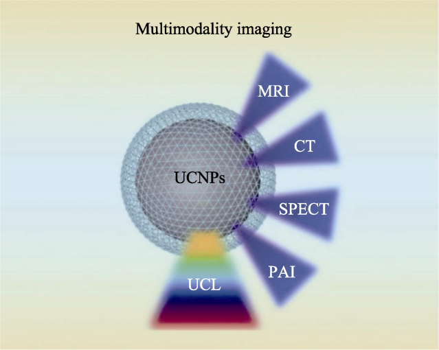
图1 UCNPs用于多模态成像
Fig. 1 Upconversion nanoparticles (UCNPs) assisted multimodal imaging
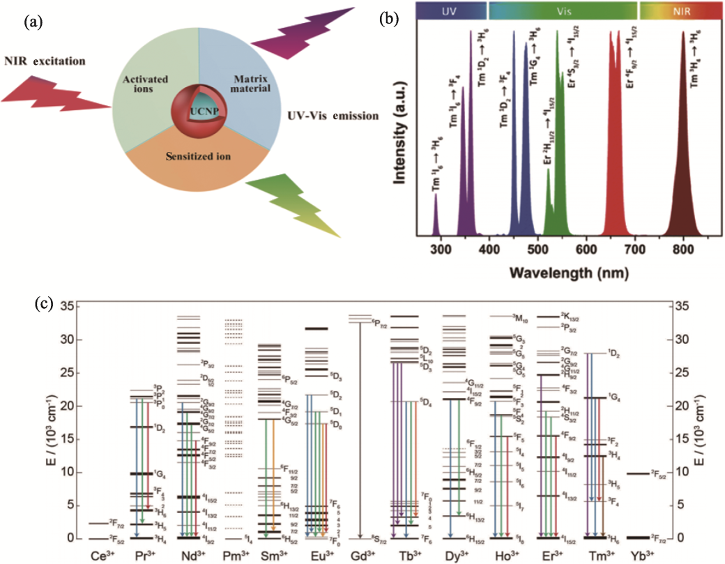
图2 UCNPs的组成、UCL和荧光发射能级图[31-32]
Fig. 2 Composition, upconversion luminescence (UCL) emissions and fluorescent energy-level diagrams of the UCNPs[31-32] (a) Schematic diagram of UCNPs; (b) Typical UCL emissions of Yb3+-Er3+ and Yb3+-Tm3+ co-doped UCNPs under 980 nm excitation[31]; (c) Partial energy-level diagrams of lanthanide ions[32]
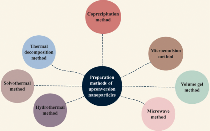
图3 UCNPs的制备方法
Fig. 3 Preparation methods of UCNPs
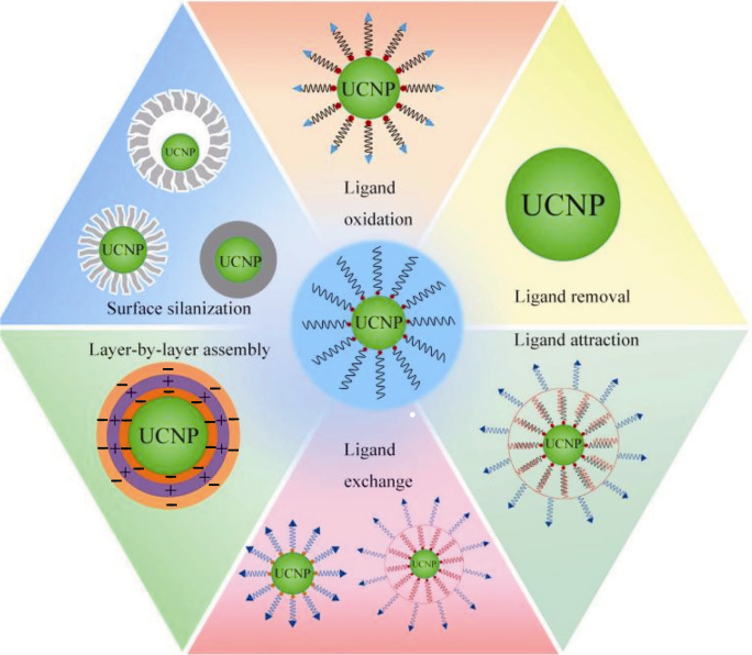
图4 UCNPs的表面修饰和改性方法
Fig. 4 Modification methods of UCNPs
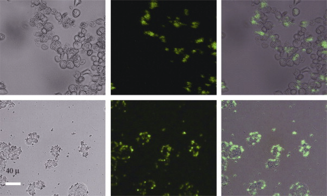
图5 PEI/NaYF4纳米颗粒用于人卵巢癌细胞和人结肠腺癌细胞的成像照片[44]
Fig. 5 Images of PEI/NaYF4 nanoparticles for human ovarian carcinoma cells and human colon adenocarcinoma cells[44] Bright field (left), confocal (middle), and superimposed (right) images of live human ovarian carcinoma cells (OVCAR3, top row) and human colon adenocarcinoma cells (HT29, bottom row). Nanoparticles were surface modified with folic acid.
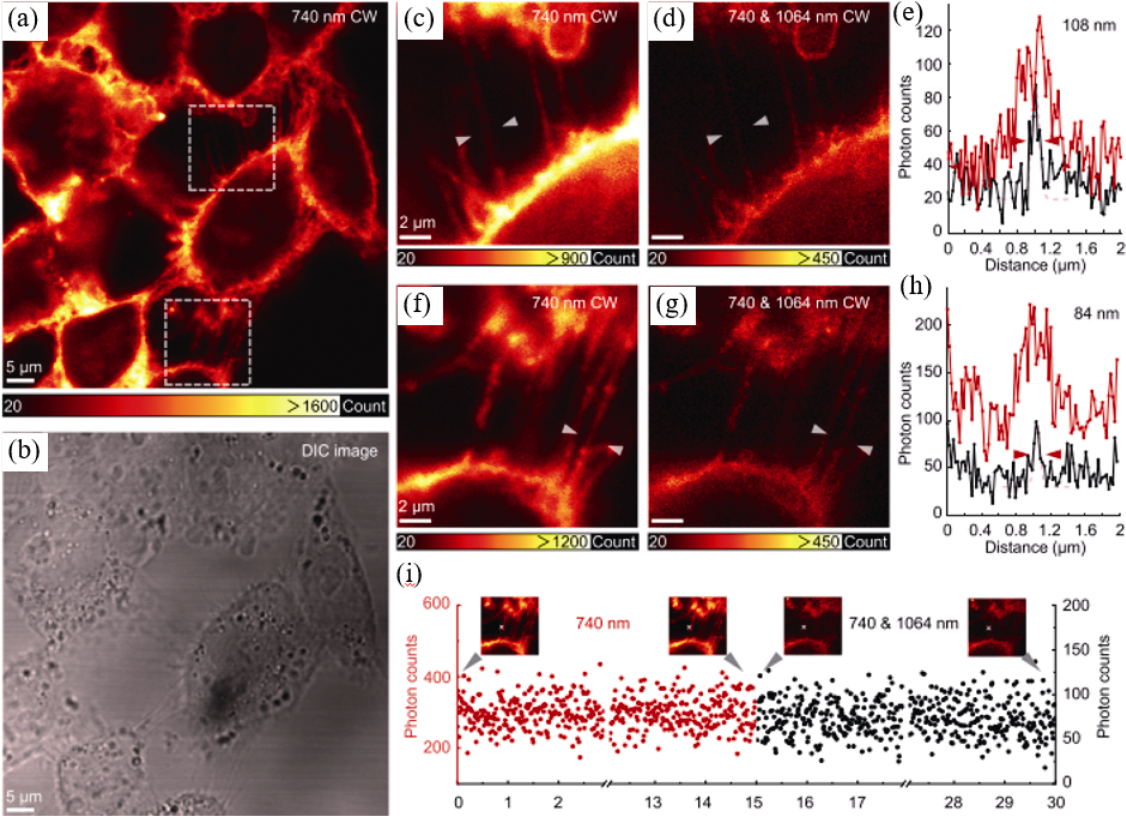
图6 免疫荧光标记细胞的超分辨成像[50]
Fig. 6 STExD (stimulated-emission induced excitation depletion) imaging of immunolabelled subcellular filaments[50] (a) Fluorescence image under 740 nm Gaussian beam excitation and (b) differential interference contrast image of actin protein in HeLa cells immunolabeled with phalloidin conjugated NaYF4: Nd nanoparticles; (c, f) Magnified areas and (d, g) super-resolution images under co-irradiation with 740 nm Gaussian beam and 1064 nm donut-shaped beam from white-dotted squares in (a); (e, h) Analyses of line profiles, indicated with white arrows in (c, d), as well as in (f, g), respectively; (i) Photon counts recorded with and without co-irradiation of 1064 nm donut-shaped beam with insets showing the corresponding images with the measured positions marked with white crosses
| Modality | Advantage | Limitation | Detection |
|---|---|---|---|
| MRI | High spatial resolution, high tissue penetration depth | Relatively low sensitivity, high cost, long imaging time, quantification | Magnetic field |
| CT | High spatial resolution | High cost | X ray |
| PET | Unlimited tissue penetration, high sensitivity, quantification | High cost | γ ray |
| SPECT | Unlimited tissue penetration, high sensitivity, quantification | Low spatial resolution | γ ray |
| PAI | High spatial resolution, unlimited tissue penetration | Low sensitivity, low tissue penetration | Ultrasonic signal |
| Fluorescence imaging | High sensitivity, multicolor imaging | Low tissue penetration, low spatial resolution | Fluorescence |
表1 纳米治疗领域的临床成像模式比较[6]
Table 1 Comparison of clinical imaging modalities used in nanotheranostice[6]
| Modality | Advantage | Limitation | Detection |
|---|---|---|---|
| MRI | High spatial resolution, high tissue penetration depth | Relatively low sensitivity, high cost, long imaging time, quantification | Magnetic field |
| CT | High spatial resolution | High cost | X ray |
| PET | Unlimited tissue penetration, high sensitivity, quantification | High cost | γ ray |
| SPECT | Unlimited tissue penetration, high sensitivity, quantification | Low spatial resolution | γ ray |
| PAI | High spatial resolution, unlimited tissue penetration | Low sensitivity, low tissue penetration | Ultrasonic signal |
| Fluorescence imaging | High sensitivity, multicolor imaging | Low tissue penetration, low spatial resolution | Fluorescence |
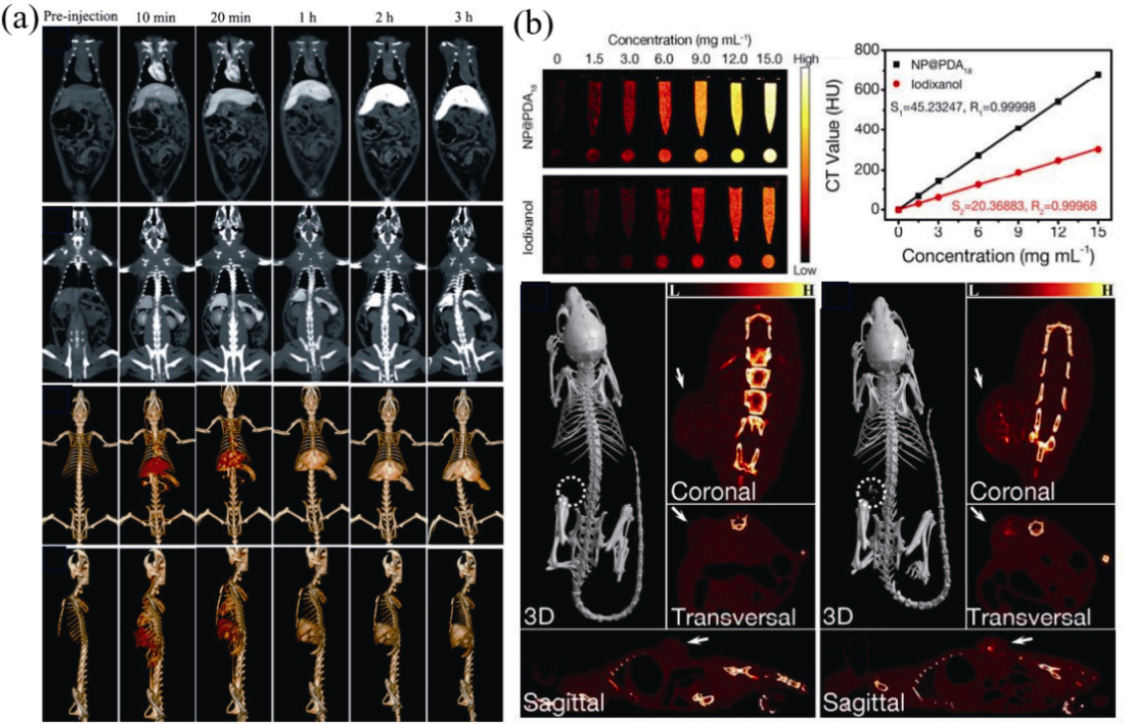
图7 含镧系元素Yb (a)和Lu (b)的UCNPs用作CT成像造影剂[56-57]
Fig. 7 UCNPs containing lanthanide elements Yb (a) and Lu (b) used as CT imaging contrast agents[56-57] (a) In vivo CT imaging after intravenous injection of 1 mL UCNPs (Yb: 70 mg·mL-1), with photos showing heart and liver, spleen and kidney, and three-dimensional CT images, respectively[56]; (b) Application of NaYF4: Nd3+@NaLuF4@PDA used for CT imaging, with photos showing X-ray images and HU values of NP@PDA18 nanocomposites and iodoxazole aqueous solution with different concentrations, and X-ray CT images of HeLa tumor bearing nude mice before and after intratumoral injection of 100 μL of NP@PDA18 solution (3 mg·mL-1)[57]
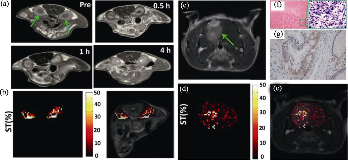
图8 UCNPs用于UCL/MRI双模态成像[66]
Fig. 8 Application of UCNPs in UCL/MRI dual-modality imaging[66] (a) In vivo T1-weighted MRI of kidneys of mice (as arrowed) before and after the intravenous administration of NaGdF4@poly-L-lysine (PLL) nanoparticles; (b) Chemical exchange saturation transfer (CEST) contrast difference map between pre/post injection following radio frequency (RF) irradiation at 3.0 μT, showing the kidney signal in color on the grayscale image to highlight the effect; (c) In vivo T1-weighted MRI of brain tumor (as arrowed) after the intravenous administration of NaGdF4@PLL nanoparticles; (d) CEST contrast difference map between pre/post injection at 3.0 μT, showing the brain ventricle signal in color on the grayscale image to highlight the effect; (e) Merged image of (c, d); (f) H&E staining of the brain tumor tissue; (g) Immunohistochemical staining of the brain tumor tissue, showing the positive expression of glial fibrillary acidic protein
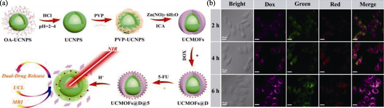
图9 NaYF4: Yb/Er@NaGdF4@金属有机框架(MOFs)@阿霉素(DOX)的制备及其UCL/MRI应用[67]
Fig. 9 Preparation and UCL/MRI application of NaYF4: Yb/Er@NaGdF4@metal-organic frameworks (MOFs) @doxorubicin (DOX)[67] (a) Schematic illustration of NaYF4: Yb/Er@NaGdF4@MOFs (UCMOFs) for dual pH-response drug release, UCL imaging and MRI; (b) Confocal fluorescence images of HeLa cells after incubation with NaYF4: Yb/Er@NaGdF4@MOFs@DOX (UCMOFs@D) for 2, 4 and 6 h at 37 ℃, with purple indicating released anticancer drug DOX, green and red indicating UCL (scale bar: 20 µm)
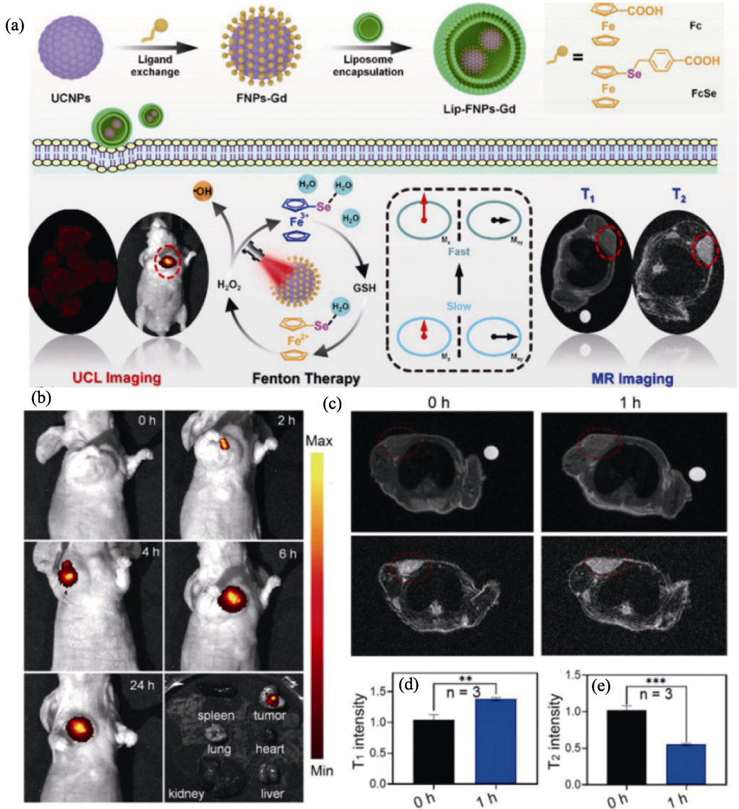
图10 Lip-FNPs-Gd纳米复合材料的制备及其在T1-T2型MRI/UCL双模态成像中的应用[68]
Fig. 10 Preparation of Lip-FNPs-Gd nanocomposite and its application in T1-T2 MRI/UCL dual-modality imaging[68] (a) Schematic illustration of preparation of Lip-FNPs-Gd nanocomposites and ferrocene/selenium-mediated applications in T1-T2 MRI/UCL multimodal imaging and NIR-promoted Fenton therapy of tumors; (b) UCL, (c) T1- and T2-weighted MRI of AGS tumor-bearing mouse for different periods of time; Corresponding T1 (d) and T2 (e) intensity analysis of tumor MRI in (c)
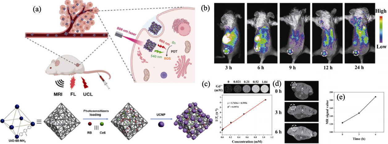
图11 双光敏剂二氢卟吩e6(Ce6)和孟加拉玫瑰红(RB)结合形成的MOF@UCNP用于MRI/UCL双模态成像[69]
Fig. 11 MOF@UCNP formed by combination of dual photosensitizers chlorin e6 (Ce6) and rose Bengal (RB) used for MRI/UCL dual-mode imaging[69] (a) Schematic illustration of fabrication process and operation for imaging-guided photodynamic therapy (dual photosensitizers of Ce6 and RB loaded into MOF nanoparticles and further combined with NaGdF4: Yb,Er@NaGdF4: Nd/Yb via electrostatic interaction to form MOF@UCNP (denoted as CR@MUP)); (b) Fluorescence images of 4T1 tumor-bearing mice at different time after intravenous injection of CR@MUP with white circles showing the tumors; (c) T1-weighted MR images and T1 relaxation curves of CR@MUP; (d) T1-weighted MR images with white circles showing the tumors; (e) Quantification analysis of MR signals of 4T1 tumor-bearing mice treated with CR@MUP
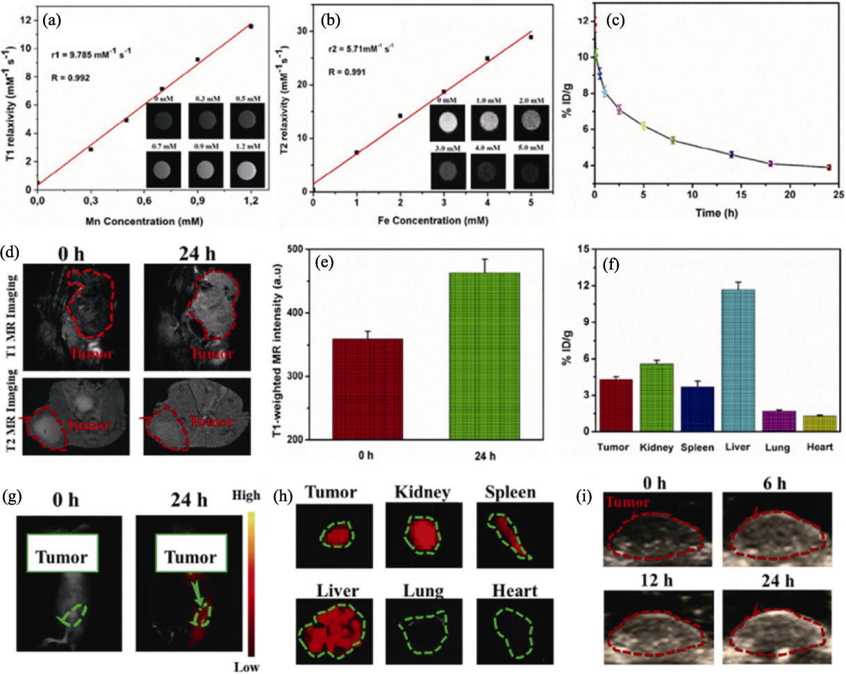
图12 MUCNPs@BPNs-Ce6复合纳米颗粒在T1、T2加权MRI中的应用[72]
Fig. 12 Application of MUCNPs@BPNs-Ce6 in T1- and T2-weighted MRI[72] (a) Linear correlation between longitudinal relativities (r1) and equivalent Mn concentrations of MUCNPs@BPNs-Ce6 at pH 6, with insets showing T1-weighted MR images of MUCNPs@BPNs-Ce6 solution at various Mn concentrations; (b) Linear correlation between longitudinal relativities (r2) and equivalent Fe concentrations of MUCNPs@BPNs-Ce6, with insets showing T2-weighted MR images of MUCNPs@BPNs-Ce6 solution at various Fe concentrations; (c) Blood time activity curve for MUCNPs@BPNs-Ce6 by measuring Fe concentration; (d) In vivo MR images of the tumor of mice before and after injection with MUCNPs@BPNs-Ce6 for 24 h; (e) T1-weighted MR signals in the tumor before and after injection with MUCNPs@BPNs-Ce6 for 24 h; (f) Biodistribution results of HeLa tumor-bearing mice before and after injection with MUCNPs@BPNs-Ce6 in MRI for 24 h; (g) In vivo fluorescence images of HeLa tumor-bearing mice before and after injection with MUCNPs@BPNs-Ce6 for 24 h; (h) Ex vivo fluorescence images of organs and tumor after injection with MUCNPs@BPNs-Ce6 for 24 h; (i) In vivo ultrasonic imaging of HeLa tumor-bearing mice after injection with MUCNPs@BPNs-Ce6 for different time
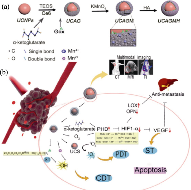
图13 NaYF4: Yb, Er@mSiO2@Ce6&α-酮戊二酸&GOx@mMnO2@HA(UCAGMH)纳米平台在乳腺肿瘤治疗中的应用[73]
Fig. 13 Schematic illustration of therapy strategy of NaYF4: Yb, Er@mSiO2@Ce6&α-ketoglutaric acid&GOx@mMnO2@HA (UCAGMH) nanoplatforms for breast tumors[73] (a) Therapeutic mechanism of UCAGMH in the mouse breast tumor model; (b) Key role that UCAGMH plays in MRI, CT and FI for therapy guidance in which HA can target CD44 receptors and induce nanoparticles to enter cells through phagocytosis
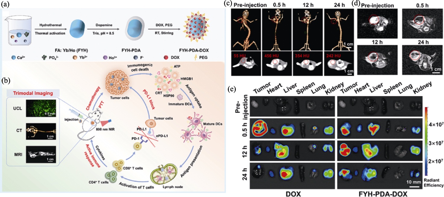
图14 FYH-PDA-DOX纳米颗粒用于UCL/CT/MRI多模态成像引导的光热治疗(PTT)-化学免疫治疗[77]
Fig. 14 FYH-PDA-DOX nanoparticles used for photothermal therapy (PTT)-chemo-immunotherapy with multimodality UCL/CT/MRI guidance[77] (a) Preparation process of FYH-PDA-DOX complex; (b) Schematic illustration using FYH-PDA-DOX for PTT-chemo-immunotherapy by combining immunogenic cell death (ICD)/ immune checkpoint blockade (ICB) with UCL/CT/MRI guidance; (c) 3D rendering of CT imaging and their corresponding coronal images of mice after injection of FYH-PDA-DOX solutions at timed intervals; (d) T2-MRI of mice before and after injection of FYH-PDA-DOX solutions; (e) Fluorescence images of excised tumors and major organs
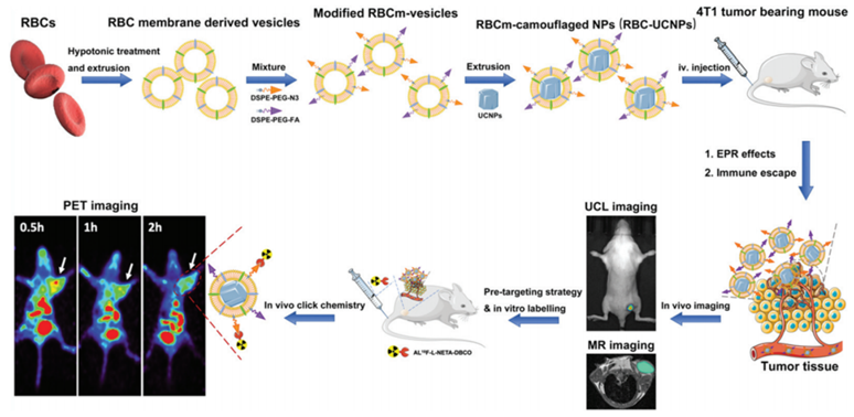
图15 RBC-UCNPs纳米颗粒的制备(上)及其在小鼠三基因乳腺癌中的MRI、UCL和PET成像(下)[79]
Fig. 15 Preparation (up row) and application of MRI, UCL and PET imaging (low row) in mice carrying three gene breast cancer of RBC-UCNPs nanoparticles[79]
| [1] | JONES K E, PATEL N G, LEVY M A, et al. Global trends in emerging infectious diseases. Nature, 2008, 451(7181): 990. |
| [2] | BRAY F, LAVERSANNE M, SUNG H, et al. Global cancer statistics 2022: GLOBOCAN estimates of incidence and mortality worldwide for 36 cancers in 185 countries. CA: A Cancer Journal for Clinicians, 2024, 74(3): 229. |
| [3] | WEN M, YU N, YI Z G, et al. On-demand phototoxicity inhibition of sensitizers and H2S-triggered in situ activation for precise therapy of colon cancer. Nano Today, 2023, 50: 101863. |
| [4] | ZHAO Y, CHEN C R, QIU Y Y, et al. Injectable fiber electronics for tumor treatment. Advanced Fiber Materials, 2022, 4(2): 246. |
| [5] | YANG J, XU L, DING Y N, et al. NIR-II-triggered composite nanofibers to simultaneously achieve intracranial hemostasis, killing superbug and residual cancer cells in brain tumor resection surgery. Advanced Fiber Materials, 2023, 5(1): 209. |
| [6] | WANG Y H, SONG S Y, ZHANG S T, et al. Stimuli-responsive nanotheranostics based on lanthanide-doped upconversion nanoparticles for cancer imaging and therapy: current advances and future challenges. Nano Today, 2019, 25: 38. |
| [7] | CHEN L, SUN X Q, CHENG K, et al. Temperature-regulating phase change fiber scaffold toward mild photothermal-chemotherapy. Advanced Fiber Materials, 2022, 4(6): 1669. |
| [8] | METTENBRINK E M, YANG W, WILHELM S. Bioimaging with upconversion nanoparticles. Advanced Photonics Research, 2022, 3(12): 2200098. |
| [9] | ANSARI A A, PARCHUR A K, THORAT N D, et al. New advances in pre-clinical diagnostic imaging perspectives of functionalized upconversion nanoparticle-based nanomedicine. Coordination Chemistry Reviews, 2021, 440: 213971. |
| [10] |
ZHU X J, SU Q Q, FENG W, et al. Anti-Stokes shift luminescent materials for bio-applications. Chemical Society Reviews, 2017, 46(4): 1025.
DOI PMID |
| [11] | SCHROTER A, HIRSCH T. Control of luminescence and interfacial properties as perspective for upconversion nanoparticles. Small, 2024, 20(14): 2306042. |
| [12] | WANG H, WANG Z H, TU Y B, et al. Homotypic targeting upconversion nano-reactor for cascade cancer starvation and deep-tissue phototherapy. Biomaterials, 2020, 235: 119765. |
| [13] |
HE S Q, SONG J, QU J L, et al. Crucial breakthrough of second near-infrared biological window fluorophores: design and synthesis toward multimodal imaging and theranostics. Chemical Society Reviews, 2018, 47(12): 4258.
DOI PMID |
| [14] | KUANG G Z, LU H T, HE S S, et al. Near-infrared light-triggered polyprodrug/siRNA loaded upconversion nanoparticles for multi-modality imaging and synergistic cancer therapy. Advanced Healthcare Materials, 2021, 10(20): e2100938. |
| [15] | AKHTAR N, CHEN C L, CHATTOPADHYAY S. PDT-active upconversion nanoheaters for targeted imaging guided combinatorial cancer phototherapies with low-power single NIR excitation. Biomaterials Advances, 2022, 141: 213117. |
| [16] |
WANG Z, LIU B, SUN Q Q, et al. Upconverted metal-organic framework Janus architecture for near-infrared and ultrasound co-enhanced high performance tumor therapy. ACS Nano, 2021, 15(7): 12342.
DOI PMID |
| [17] | 彭皓, 杨方, 杜慧, 等. 基于Er3+掺杂上转换纳米粒子的生物成像研究进展. 分析化学, 2021, 49(7): 1106. |
| [18] | GAO W, ZHANG C X, HAN Q Y, et al. Enhancing red upconversion emission of Ho3+ in a single NaYbF4: Ho3+ microdisk through building different core-shell structures. Journal of Luminescence, 2022, 241: 118501. |
| [19] | LIU S B, AN Z C, ZHOU B. Optical multiplexing of upconversion in nanoparticles towards emerging applications. Chemical Engineering Journal, 2023, 452: 139649. |
| [20] | CHEN H, DING B B, MA P A, et al. Recent progress in upconversion nanomaterials for emerging optical biological applications. Advanced Drug Delivery Reviews, 2022, 188: 114414. |
| [21] | RAFIQUE R, KAILASA S K, PARK T J. Recent advances of upconversion nanoparticles in theranostics and bioimaging applications. TrAC Trends in Analytical Chemistry, 2019, 120: 115646. |
| [22] | HONG E L, LIU L M, BAI L M, et al. Control synthesis, subtle surface modification of rare-earth-doped upconversion nanoparticles and their applications in cancer diagnosis and treatment. Materials Science & Engineering C-Materials for Biological Applications, 2019, 105: 110097. |
| [23] | BAO W E, LIU M, MENG J Q, et al. MOFs-based nanoagent enables dual mitochondrial damage in synergistic antitumor therapy via oxidative stress and calcium overload. Nature Communications, 2021, 12(1): 6399. |
| [24] | LI Y M, LI Y M, BAI Y D, et al. Activating ultralow upconversion nanothermometry in neodymium sublattice for heart tissue imaging rapid-responsive. Talanta, 2023, 264: 124764. |
| [25] | ZHANG Z, CHEN Y M, ZHANG Y. Self-assembly of upconversion nanoparticles based materials and their emerging applications. Small, 2022, 18(9): 2103241. |
| [26] | GAO W, CHENG X T, XING Y, et al. Enhancement of red upconversion emission intensity of Ho3+ ions in NaLuF4: Yb3+/Ho3+/Ce3+@NaLuF4 core-shell nanoparticles. Journal of Rare Earths, 2022, 40(4): 517. |
| [27] | LIU S B, YAN L, LI Q Q, et al. Tri-channel photon emission of lanthanides in lithium-sublattice core-shell nanostructures for multiple anti-counterfeiting. Chemical Engineering Journal, 2020, 397: 125451. |
| [28] |
CHEN B, WANG F. Combating concentration quenching in upconversion nanoparticles. Accounts of Chemical Research, 2020, 53(2): 358.
DOI PMID |
| [29] |
HLAVÁČEK A, FARKA Z, MICKERT M J, et al. Bioconjugates of photon-upconversion nanoparticles for cancer biomarker detection and imaging. Nature Protocols, 2022, 17(4): 1028.
DOI PMID |
| [30] | AUZEL F. Upconversion and anti-stokes processes with f and d ions in solids. ChemInform, 2004, 104(1): 139. |
| [31] |
DONG H, SUN L D, YAN C H. Energy transfer in lanthanide upconversion studies for extended optical applications. Chemical Society Reviews, 2015, 44(6): 1608.
DOI PMID |
| [32] |
WANG Y, ZHENG K Z, SONG S Y, et al. Remote manipulation of upconversion luminescence. Chemical Society Reviews, 2018, 47(17): 6473.
DOI PMID |
| [33] | HLAVÁČEK A, KŘIVÁNKOVÁ J, PIZÚROVÁ N, et al. Photon-upconversion barcode for monitoring an enzymatic reaction with a fluorescence reporter in droplet microfluidics. The Analyst, 2020, 145(23): 7718. |
| [34] |
WU S W, HAN G, MILLIRON D J, et al. Non-blinking and photostable upconverted luminescence from single lanthanide-doped nanocrystals. Proceedings of the National Academy of Sciences of the United States of America, 2009, 106(27): 10917.
DOI PMID |
| [35] |
GNACH A, LIPINSKI T, BEDNARKIEWICZ A, et al. Upconverting nanoparticles: assessing the toxicity. Chemical Society Reviews, 2015, 44(6): 1561.
DOI PMID |
| [36] | SABU A, LIN J Y, DOONG R A, et al. Prospects of an engineered tumor-targeted nanotheranostic platform based on NIR-responsive upconversion nanoparticles. Materials Advances, 2021, 2(22): 7101. |
| [37] | SUN C N, GRADZIELSKI M. Advances in fluorescence sensing enabled by lanthanide-doped upconversion nanophosphors. Advances in Colloid and Interface Science, 2022, 300: 102579. |
| [38] | 李静芝, 高志贤, 李双, 等. 上转换纳米颗粒的发光机理、制备及生物应用进展. 材料导报, 2022, 36(14): 98. |
| [39] |
ZHANG L, JIN D Y, STENZEL M H. Polymer-functionalized upconversion nanoparticles for light/imaging-guided drug delivery. Biomacromolecules, 2021, 22(8): 3168.
DOI PMID |
| [40] | TESCH A, RÖDER R, ZAPF M, et al. Paramagnetic, NIR-luminescent Nd3+- and Gd3+-doped fluorapatite as contrast agent for multimodal biomedical imaging. Journal of the American Ceramic Society, 2018, 101(10): 4441. |
| [41] |
GENERALOVA A N, CHICHKOV B N, KHAYDUKOV E V. Multicomponent nanocrystals with anti-Stokes luminescence as contrast agents for modern imaging techniques. Advances in Colloid and Interface Science, 2017, 245: 1.
DOI PMID |
| [42] | YE S H, ZHANG W J, SHEN Y, et al. Simultaneous imaging and photodynamic-enhanced photothermal inhibition of cancer cells using a multifunctional system combining indocyanine green and polydopamine-preloaded upconversion luminescent nanoparticles. Macromolecular Rapid Communications, 2023, 44(20): 2300298. |
| [43] | ZHAO J, DI Z H, LI L L. Spatiotemporally selective molecular imaging via upconversion luminescence-controlled, DNA-based biosensor technology. Angewandte Chemie International Edition, 2022, 61(32): e202204277. |
| [44] |
CHATTERJEE D K, RUFAIHAH A J, ZHANG Y. Upconversion fluorescence imaging of cells and small animals using lanthanide doped nanocrystals. Biomaterials, 2008, 29(7): 937.
DOI PMID |
| [45] | CHEN S, WEITEMIER A Z, ZENG X, et al. Near-infrared deep brain stimulation via upconversion nanoparticle-mediated optogenetics. Science, 2018, 359(6376): 679. |
| [46] | TIAN G, GU Z J, ZHOU L J, et al. Mn2+ dopant-controlled synthesis of NaYF4: Yb/Er upconversion nanoparticles for in vivo imaging and drug delivery. Advanced Materials, 2012, 24(9): 1226. |
| [47] | ZHAN Q Q, QIAN J, LIANG H J, et al. Using 915 nm laser excited Tm3+/Er3+/Ho3+-doped NaYbF4 upconversion nanoparticles for in vitro and deeper in vivo bioimaging without overheating irradiation. ACS Nano, 2011, 5(5): 3744. |
| [48] | WANG Y F, LIU G Y, SUN L D, et al. Nd3+-sensitized upconversion nanophosphors: efficient in vivo bioimaging probes with minimized heating effect. ACS Nano, 2013, 7(8): 7200. |
| [49] | LIU B, CHEN Y Y, LI C X, et al. Poly(acrylic acid) modification of Nd3+-sensitized upconversion nanophosphors for highly efficient UCL imaging and pH-responsive drug delivery. Advanced Functional Materials, 2015, 25(29): 4717. |
| [50] |
GUO X, PU R, ZHU Z M, et al. Achieving low-power single-wavelength-pair nanoscopy with NIR-II continuous-wave laser for multi-chromatic probes. Nature Communications, 2022, 13(1): 2843.
DOI PMID |
| [51] | SHEN J W, YANG C X, DONG L X, et al. Incorporation of computed tomography and magnetic resonance imaging function into NaYF4: Yb/Tm upconversion nanoparticles for in vivo trimodal bioimaging. Analytical Chemistry, 2013, 85(24): 12166. |
| [52] | FENG Y, CHEN H D, MA L N, et al. Surfactant-free aqueous synthesis of novel Ba2GdF7: Yb3+, Er3+@PEG upconversion nanoparticles for in vivo trimodality imaging. ACS Applied Materials & Interfaces, 2017, 9(17): 15096. |
| [53] | YANG X, SONG R T, GONG X C, et al. Multi-shell structured nanomaterials with strong red upconversion emission for trimodal biomedical imaging. Ceramics International, 2024, 50(1): 1601. |
| [54] |
LIU J, ZHANG J, HUANG F, et al. X-ray and NIR light dual-triggered mesoporous upconversion nanophosphor/Bi heterojunction radiosensitizer for highly efficient tumor ablation. Acta Biomaterialia, 2020, 113: 570.
DOI PMID |
| [55] |
XING H Y, BU W B, ZHANG S J, et al. Multifunctional nanoprobes for upconversion fluorescence, MR and CT trimodal imaging. Biomaterials, 2012, 33(4): 1079.
DOI PMID |
| [56] | LIU Y L, AI K L, LIU J H, et al. A high-performance ytterbium-based nanoparticulate contrast agent for in vivo X-ray computed tomography imaging. Angewandte Chemie International Edition, 2012, 51(6): 1437. |
| [57] | DAI Y, YANG D P, YU D P, et al. Mussel-inspired polydopamine-coated lanthanide nanoparticles for NIR-II/CT dual imaging and photothermal therapy. ACS Applied Materials & Interfaces, 2017, 9(32): 26674. |
| [58] | WANG J, GUO H Y, WANG H, et al. NaGdF4-based magnetic resonance nanoprobes for qualitative inflammation imaging in glioma: hot or cold? Chemical Engineering Journal, 2024, 479: 147916. |
| [59] | LING B, WANG Y G, MI R, et al. Multimodal imaging and synergetic chemodynamic/photodynamic therapy achieved using an NaGdF4, Yb, Er@NaGdF4, Yb, Tm@NaYF4@Fe-MOFs nanocomposite. Chemistry - An Asian Journal, 2022, 17(14): e202200161. |
| [60] | JIANG Z L, XIA B, REN F, et al. Boosting vascular imaging-performance and systemic biosafety of ultra-small NaGdF4 nanoparticles via surface engineering with rationally designed novel hydrophilic block co-polymer. Small Methods, 2022, 6(3): 2101145. |
| [61] | JU Q, TU D T, LIU Y S, et al. Amine-functionalized lanthanide-doped KGdF4 nanocrystals as potential optical/magnetic multimodal bioprobes. Journal of the American Chemical Society, 2012, 134(2): 1323. |
| [62] |
YANG D M, DAI Y L, LIU J H, et al. Ultra-small BaGdF5-based upconversion nanoparticles as drug carriers and multimodal imaging probes. Biomaterials, 2014, 35(6): 2011.
DOI PMID |
| [63] | ZHU G N, CHEN L P, ZENG F X, et al. GdVO4: Eu3+, Bi3+ nanoparticles as a contrast agent for MRI and luminescence bioimaging. ACS Omega, 2019, 4(14): 15806. |
| [64] | BIJU S, GALLO J, BAÑOBRE-LÓPEZ M, et al. A magnetic chameleon: biocompatible lanthanide fluoride nanoparticles with magnetic field dependent tunable contrast properties as a versatile contrast agent for low to ultrahigh field MRI and optical imaging in biological window. Chemistry - A European Journal, 2018, 24(29): 7277. |
| [65] | LI C X, YANG D M, MA P A, et al. Multifunctional upconversion mesoporous silica nanostructures for dual modal imaging and in vivo drug delivery. Small, 2013, 9(24): 4150. |
| [66] |
NI D L, SHEN Z W, ZHANG J W, et al. Integrating anatomic and functional dual-mode magnetic resonance imaging: design and applicability of a bifunctional contrast agent. ACS Nano, 2016, 10(3): 3783.
DOI PMID |
| [67] |
LING D P, LI H H, XI W S, et al. Heterodimers made of metal-organic frameworks and upconversion nanoparticles for bioimaging and pH-responsive dual-drug delivery. Journal of Materials Chemistry B, 2020, 8(6): 1316.
DOI PMID |
| [68] |
ZHOU T, ZHANG S Y, ZHANG L, et al. Redox ferrocenylseleno compounds modulate longitudinal and transverse relaxation times of FNPs-Gd MRI contrast agents for multimodal imaging and photo-Fenton therapy. Acta Biomaterialia, 2023, 164: 496.
DOI PMID |
| [69] | LI Z K, QIAO X, HE G H, et al. Core-satellite metal-organic framework@upconversion nanoparticle superstructures via electrostatic self-assembly for efficient photodynamic theranostics. Nano Research, 2020, 13(12): 3377. |
| [70] | YANG D, XU J T, YANG G X, et al. Metal-organic frameworks join hands to create an anti-cancer nanoplatform based on 808 nm light driving up-conversion nanoparticles. Chemical Engineering Journal, 2018, 344: 363. |
| [71] |
KORETSKY A P, SILVA A C. Manganese-enhanced magnetic resonance imaging (MEMRI). NMR in Biomedicine, 2004, 17(8): 527.
PMID |
| [72] | ZHANG Q C, WANG W T, ZHANG M, et al. A theranostic nanocomposite with integrated black phosphorus nanosheet, Fe3O4@MnO2-doped upconversion nanoparticles and chlorin for simultaneous multimodal imaging, highly efficient photodynamic and photothermal therapy. Chemical Engineering Journal, 2020, 391: 123525. |
| [73] | YAN J H, SHAO K, WU L J, et al. Upconversion-nanoparticle-based smart drug-delivery platforms for multimodal imaging-guided cancer therapies. ACS Applied Nano Materials, 2022, 5(10): 15473. |
| [74] | XIAO Q F, ZHENG X P, BU W B, et al. A core/satellite multifunctional nanotheranostic for in vivo imaging and tumor eradication by radiation/photothermal synergistic therapy. Journal of the American Chemical Society, 2013, 135(35): 13041. |
| [75] |
LV R C, YANG P P, HE F, et al. A yolk-like multifunctional platform for multimodal imaging and synergistic therapy triggered by a single near-infrared light. ACS Nano, 2015, 9(2): 1630.
DOI PMID |
| [76] | XIA A, CHEN M, GAO Y, et al. Gd3+ complex-modified NaLuF4-based upconversion nanophosphors for trimodality imaging of NIR-to-NIR upconversion luminescence, X-ray computed tomography and magnetic resonance. Biomaterials, 2012, 33(21): 5394. |
| [77] | GU M Q, ZHANG L L, HAO L Y, et al. Upconversion nanoplatform enables multimodal imaging and combinatorial immunotherapy for synergistic tumor treatment and monitoring. ACS Applied Materials & Interfaces, 2023, 15(18): 21766. |
| [78] |
SUN Y, ZHU X J, PENG J J, et al. Core-shell lanthanide upconversion nanophosphors as four-modal probes for tumor angiogenesis imaging. ACS Nano, 2013, 7(12): 11290.
DOI PMID |
| [79] |
LI M T, FANG H Y, LIU Q Y, et al. Red blood cell membrane-coated upconversion nanoparticles for pretargeted multimodality imaging of triple-negative breast cancer. Biomaterials Science, 2020, 8(7): 1802.
DOI PMID |
| [1] | 陈明俊, 缪洪康, 肖英俊, 邓建波, 张翔, 赵九蓬, 李垚. 光-热双响应材料研究进展: 从设计策略到智能窗应用[J]. 无机材料学报, 2026, 41(6): 723-738. |
| [2] | 宋坤洁, 解荣军. 机器学习驱动新型发光材料的研究进展[J]. 无机材料学报, 2026, 41(6): 689-703. |
| [3] | 胡钰晴, 朱一新, 乐先浩, 万青. 钽酸锂晶圆减薄技术及其热释电红外探测器应用进展[J]. 无机材料学报, 2026, 41(6): 764-774. |
| [4] | 刘春帆, 陈科, 葛芳芳, 黄庆. 核用耐铅铋腐蚀涂层的研究进展[J]. 无机材料学报, 2026, 41(6): 775-786. |
| [5] | 胡扬, 谢敏, 张筱怡, 李想, 郭新伟, 姜南, 周文瀚, 张胜利, 曾海波. 计算与数据驱动环保型发光材料的研究进展[J]. 无机材料学报, 2026, 41(6): 704-722. |
| [6] | 王俊卜, 黄泽皑, 杨茗凯, 蒙颖, 周明炜, 周莹. 甲烷转化用抗积碳催化材料研究进展[J]. 无机材料学报, 2026, 41(6): 739-750. |
| [7] | 王金文, 杨振, 周欢, 夏丹, 杨磊. 可注射无机材料及其生物医学应用[J]. 无机材料学报, 2026, 41(6): 751-763. |
| [8] | 李涵涛, 沈强, 罗国强, 王雪飞, 高明, 陈晨. 机械球磨法调控硅基负极材料结构与性能的研究进展[J]. 无机材料学报, 2026, 41(5): 561-572. |
| [9] | 解陈一, 缪花明, 张蔚然, 刘荣军, 王衍飞, 李端. 理论计算在高熵陶瓷领域的研究进展[J]. 无机材料学报, 2026, 41(5): 545-560. |
| [10] | 李璇, 叶奎材, 冯佳音, 邱家军, 钱文昊, 邢敏. 钛基牙种植体表面改性促进软组织封闭的研究进展[J]. 无机材料学报, 2026, 41(4): 432-444. |
| [11] | 彭德招, 李瑞, 王文鸿, 王梓瑞, 章志珍. 钠氯化物固态电解质研究进展[J]. 无机材料学报, 2026, 41(4): 409-420. |
| [12] | 陈坤, 姜勇刚, 冯军宗, 李良军, 胡艺洁, 冯坚. 锆酸镧多孔隔热材料研究进展[J]. 无机材料学报, 2026, 41(4): 421-431. |
| [13] | 韦连金, 齐志杰, 汪信, 朱俊武, 付永胜. 纳米金刚石改性及其在电催化氧还原反应中的应用[J]. 无机材料学报, 2026, 41(3): 273-288. |
| [14] | 刘占一, 李勉, 欧阳晓平, 柴之芳, 黄庆. 干法后处理熔盐中Sr/Cs去除方法的研究进展[J]. 无机材料学报, 2026, 41(2): 150-158. |
| [15] | 孙炼, 张磊磊, 薛泽旭, 吴坤, 陈晔, 李志远, 王鲁凯, 王尊刚. 面向辐射探测应用的零维金属卤化物闪烁体研究进展[J]. 无机材料学报, 2026, 41(2): 159-176. |
| 阅读次数 | ||||||
|
全文 |
|
|||||
|
摘要 |
|
|||||