无机材料学报 ›› 2014, Vol. 29 ›› Issue (10): 1009-1017.DOI: 10.15541/jim20140010 CSTR: 32189.14.10.15541/jim20140010
• • 下一篇
李 波, 徐文峰, 廖晓玲
收稿日期:2014-01-06
修回日期:2014-02-27
出版日期:2014-10-20
网络出版日期:2014-09-22
作者简介:李 波(1980–), 男, 博士, 副教授. E-mail: leewave@126.com
基金资助:LI Bo, XU Wen-Feng, LIAO Xiao-Ling
Received:2014-01-06
Revised:2014-02-27
Published:2014-10-20
Online:2014-09-22
About author:LI Bo. E-mail: leewave@126.com
Supported by:摘要:
磷酸钙微球具有良好的渗透性、高的比表面积、低致密度和较好的力学性能,在分离、催化、传感、组织工程和药物释放等方面均有应用。本文综述了近年来磷酸钙陶瓷微球在组织工程和药物释放等骨修复相关领域的研究进展, 介绍了实心、多孔、空心和花瓣状等四种不同结构磷酸钙陶瓷微球制备方法以及在骨修复领域中的应用, 并归纳总结了各类微球具有的优缺点和改进的方向, 为骨修复用磷酸钙微球的设计和制备提供较系统的参考。
中图分类号:
李 波, 徐文峰, 廖晓玲. 磷酸钙微球骨修复材料研究进展[J]. 无机材料学报, 2014, 29(10): 1009-1017.
LI Bo, XU Wen-Feng, LIAO Xiao-Ling. Research Progress in Calcium Phosphate Microspheres for Bone Defect Repair[J]. Journal of Inorganic Materials, 2014, 29(10): 1009-1017.
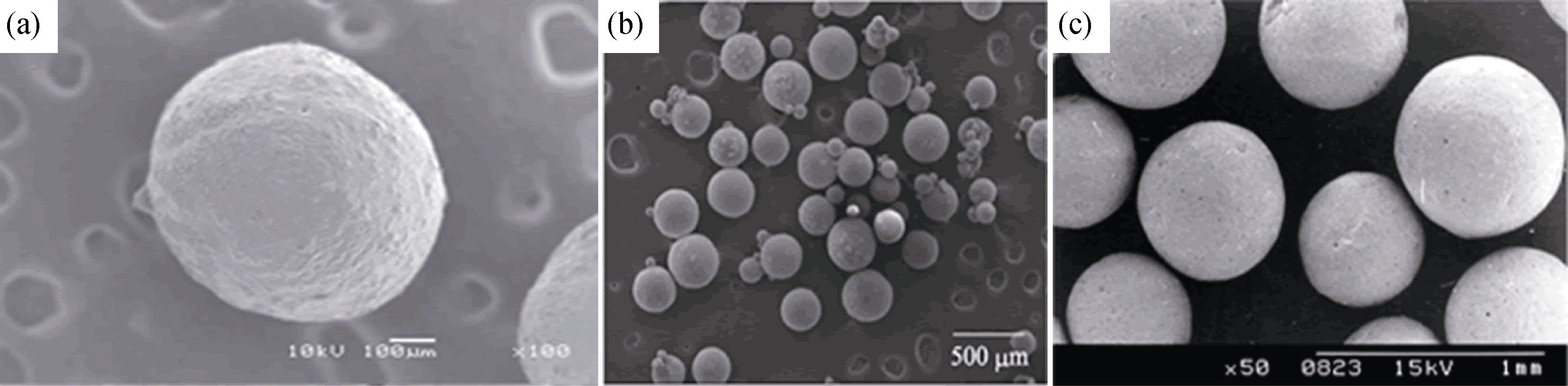
图1 几种典型的CaP陶瓷实心微球[10, 12-13]
Fig. 1 Images of typical calcium phosphate solid microspheres (a) β-TCP microsphere prepared with nitrogen liquid freeze drying method[10]; (b) HA microsphere prepared with CMCS and gel as binder[12]; (c) HA microsphere prepared with emulsion method[13]
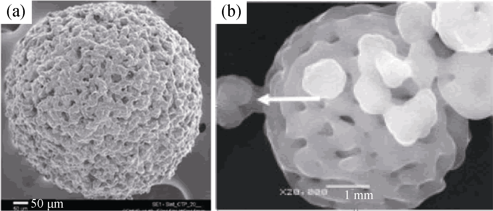
图2 两种典型的多孔CaP微球[18, 23]
Fig. 2 Two typical porous CaP microspheres[18, 23] (a) HA microsphere prepared with alginate salt gel process[18], (b) HA microsphere prepared with spray drying method[23]
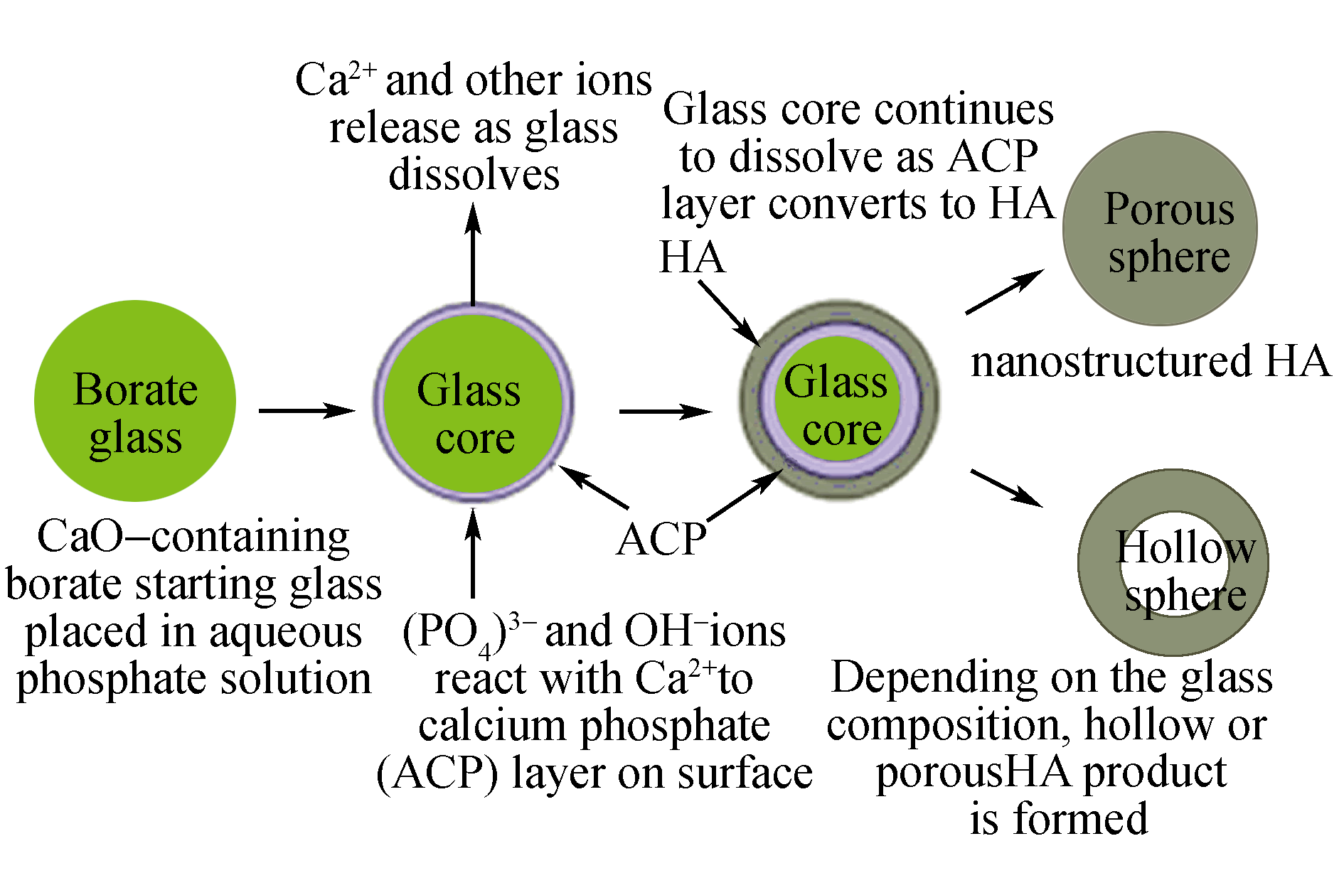
图3 以生物玻璃微球为硬模板的方式制备的CaP陶瓷空心微球工艺流程示意图[27]
Fig. 3 Scheme of calcium phosphate hollow microsphere with bioglass as hard template[27]
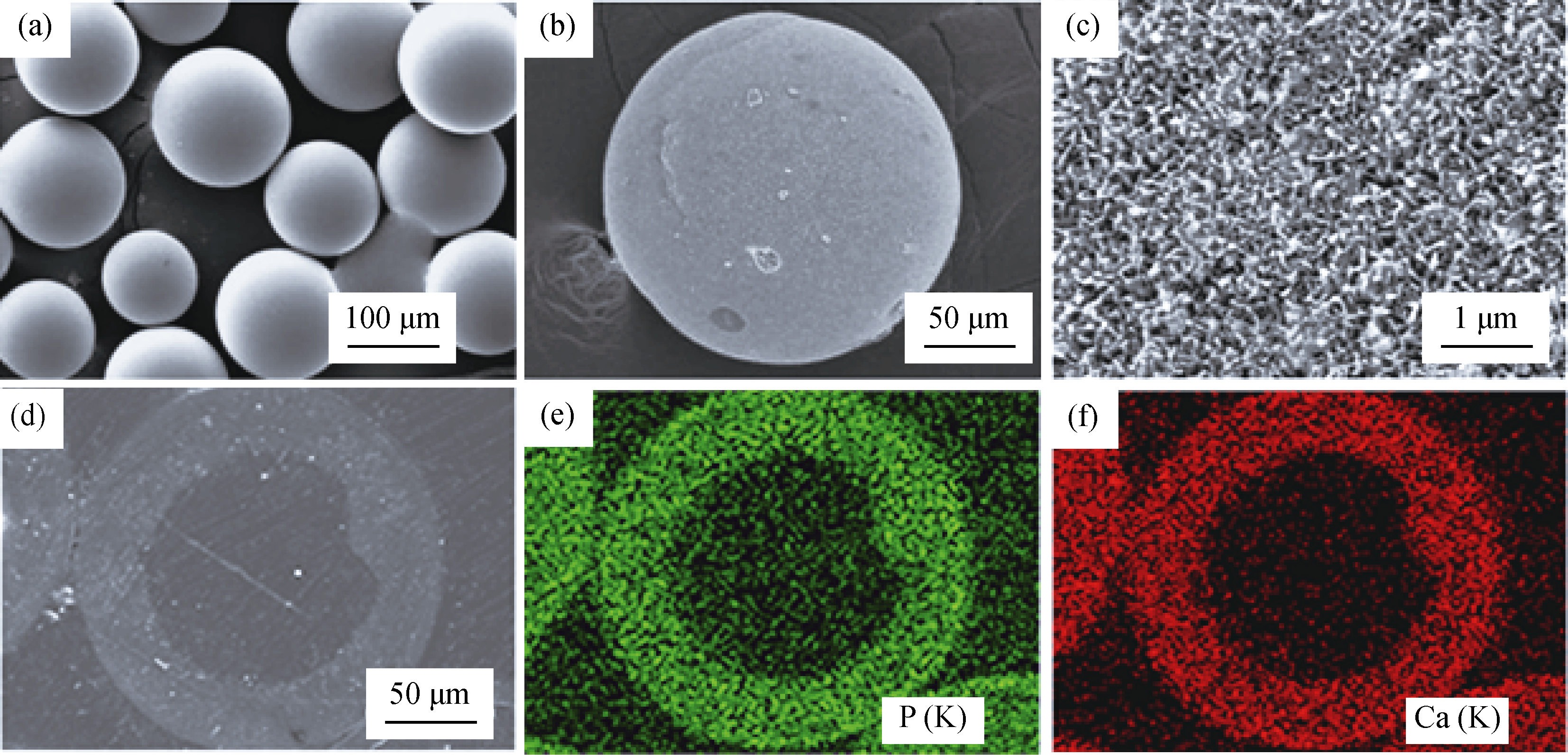
图4 以玻璃微球为硬模板制备的空心CaP陶瓷时, 作为模板用玻璃微球形貌(a), 空心HA微球外表面形貌(b),空心HA表面放大形貌(c), HA空心微球抛光纵切面背散射SEM观察到的形貌(d), X射线能谱显示图片(d)的含P(e)和Ca(f)元素分布 [27]
Fig. 4 SEM images of hollow HA microsphere with glass as hard template. (a) starting glass microspheres as hard template, (b) external surface of hollow HA microsphere, (c) external surface of hollow HA microsphere at high magnification. (d) SEM image in back-scattered mode of a polished cross section of a hollow hydroxyapatite microsphere, (e) and (f) X-ray maps of Ca(K) and P(K) across the planar section shown in (d)[27]
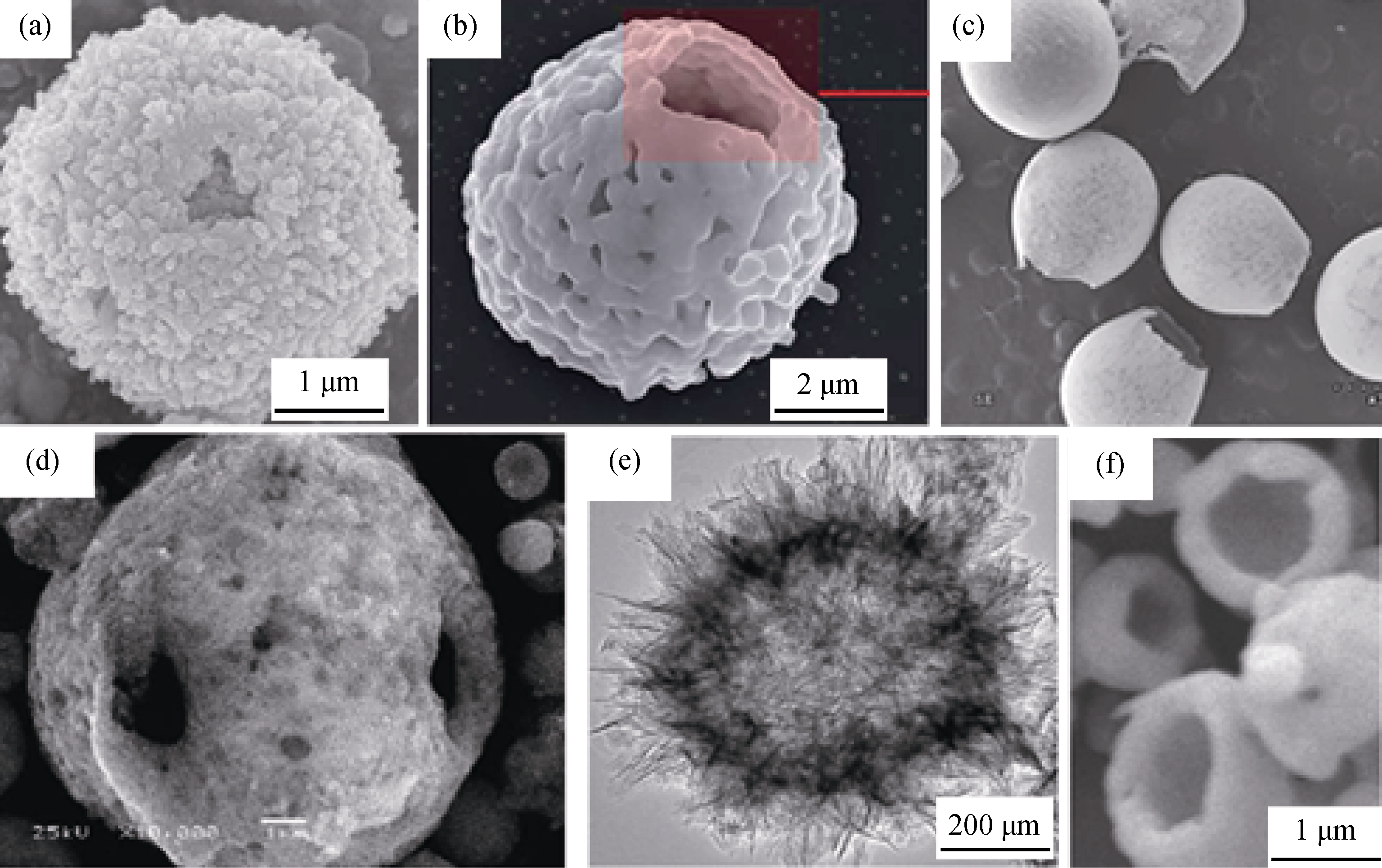
图5 分别以(a) CaCO3/Fe3O4硬模板法[32]、(b)酵母菌生物模板法[33]、(c) DCM乳液法[34]、(d) 喷雾干燥法[35]、(e) 微波水热法[37]、和(f) 电喷涂法[38]制备的几种典型的空心CaP陶瓷微球
Fig. 5 Six typical hollow calcium phosphate microspheres prepared with (a) CaCO3/Fe3O4 as hard template[32], (b) yeast as bio- template[33], (c) DCM emulsion method[34], (d) spray drying method[35], (e) microwave-hydrothermal method[37], and (f) electrosprayed method[38], respectively
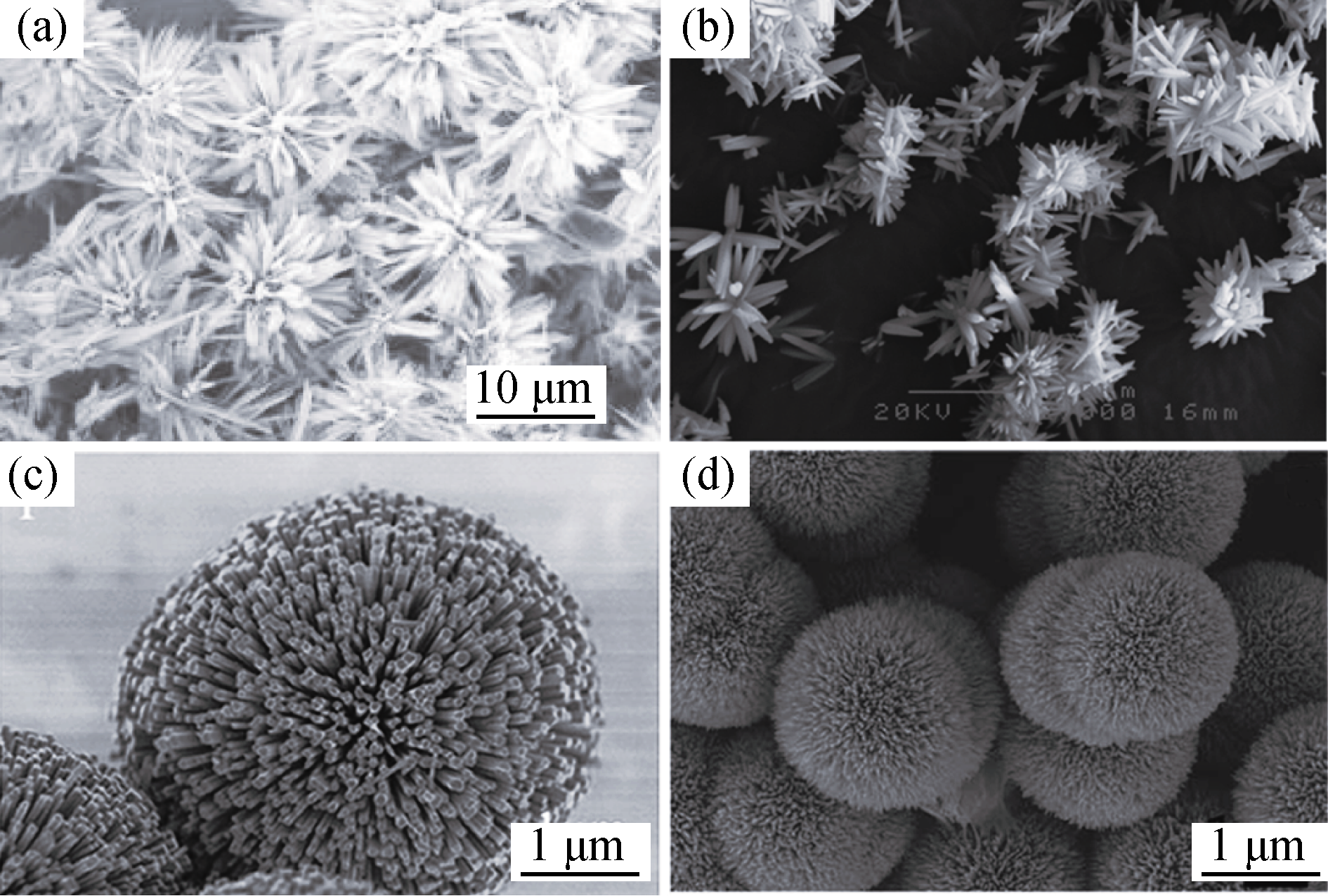
图6 4种典型的花瓣状CaP陶瓷微球SEM照片, 分别是以 (a) 酒石酸氢钾[39]、(b) EDTA[40]、(c) 氟取代结合EDTA [41]和(d) 柠檬酸[42]等为模板制备而成
Fig. 6 Four typical flower-like microspheres prepared with potassium hydrogen tartrate (a)[39], EDTA[40] (b), F- substitution combined with EDTA (c) and citric acid[41] (d) as template, respectively[42]
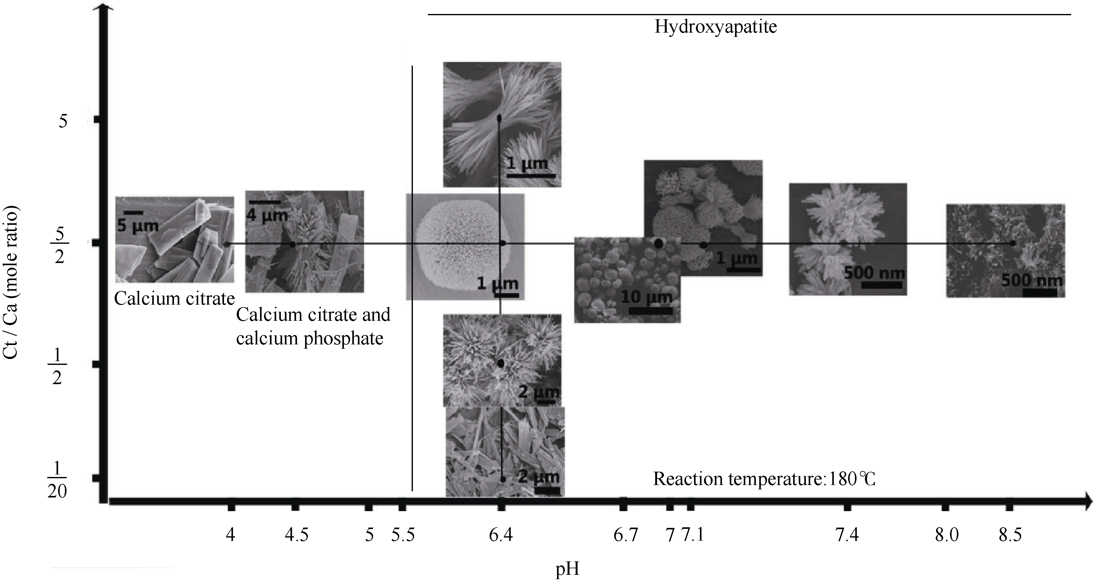
图7 以柠檬酸为模板制备CaP陶瓷微球随pH和Ct/Ca比值的形态变化图谱[42]
Fig. 7 Morphology variation of HA crystals from solution at different pH and Ct/Ca mole ratio[42]
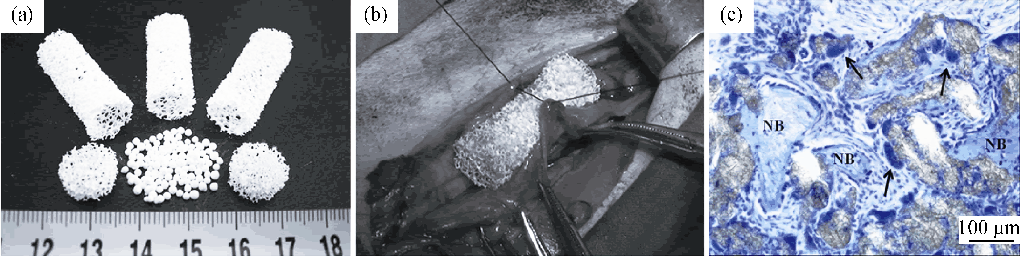
图8 HA微球与管状多孔HA支架及多孔圆片构建骨修复体(a)和腹膜植入(b)[45], 肌内植入3个月(c)后甲苯胺蓝染色组织切片[46]
Fig. 8 Images of (a) the HA spherules, porous HA tubes, and HA disks fabricated to assemble the novel scaffolds, (b) digital photo shows the implantation of the porous scaffolds at peritoneum pocket[45], (c) photographs of toluidine blue stained spherulite HA-positive assemble scaffold after intramuscular implantation for 3 months[46]. NB represents new bone
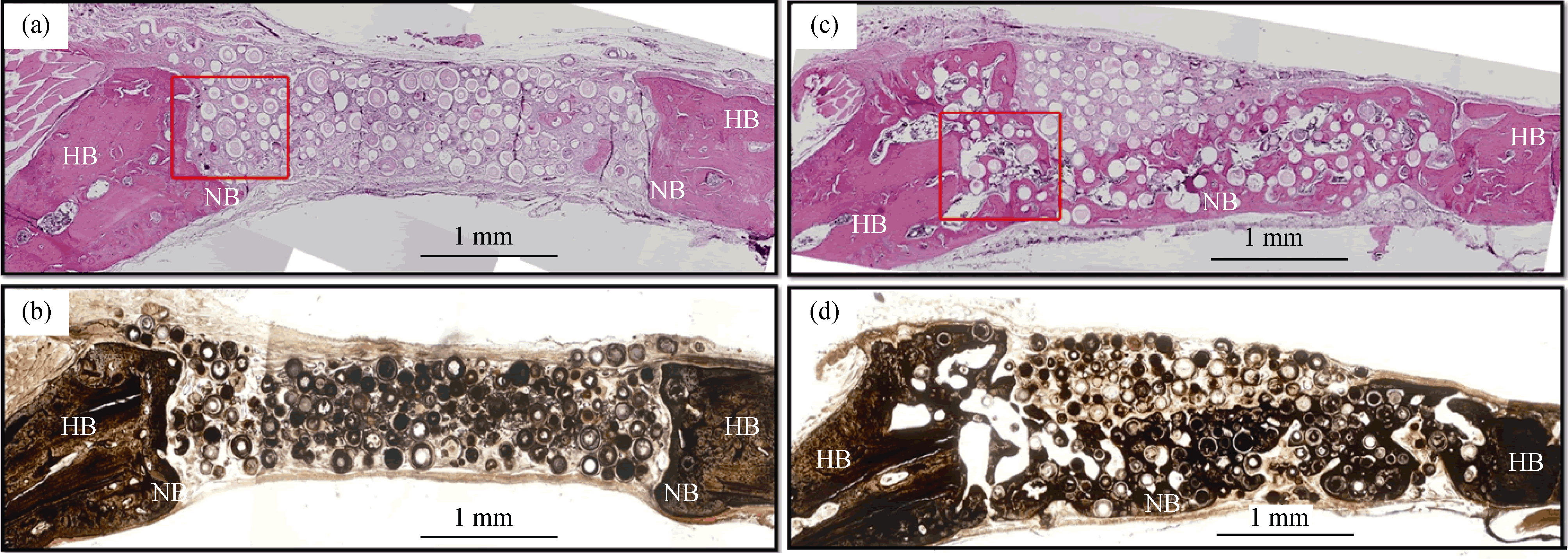
图9 空心CaP陶瓷微球修复大鼠颅骨6w后HE染色(a, c)和von Koss染色(b, d)组织切片, 其中(a, b)为空白对照微球, (c, d)为载1 μg BMP2微球[56]
Fig. 9 (a, c) H&E and (b, d) von Kossa stained sections of rat calvarial defects implanted for 6 weeks with (a, b) as-prepared hollow HA microspheres (without BMP2) and (c, d) hollow HA microspheres loaded with BMP2 (1 μg per defect). HB represents host (old) bone; NB represents new bone[56]
| [1] | WANG H, LEEUWENBURGH S C G, LI Y, et al. The use of micro- and nano-spheres as functional components for bone tissue regeneration. Tissue. Eng. Part B Rev., 2012, 18(1): 24-39. |
| [2] | LIU X, JIN X, MA P X. Nanofibrous hollow microspheres self-assembled from star-shaped polymers as injectable cell carriers for knee repair. Nat. Mater., 2011, 10(5): 398-406. |
| [3] | FISHER M B, MAUCK R L. Tissue engineering and regenerative medicine: recent innovations and the transition to translation. Tissue Engineering Part B-Reviews, 2013, 19(1): 1-13. |
| [4] | HONG Y, FAN H, LI B, et al. Fabrication, biological effects, and medical applications of calcium phosphate nanoceramics. Mat. Sci. Eng. R., 2010, 70(3-6): 225-242. |
| [5] | SADAT-SHOJAI M, KHORASANI M T, DINPANAH- KHOSHDARGI E, et al. Synthesis methods for nanosized hydroxyapatite with diverse structures. Acta Biomater., 2013, 9(8): 7591-7621. |
| [6] | CHRISTENSON E M, ANSETH K S, VAN DEN BEUCKEN L, et al. Nanobiomaterial applications in orthopedics. J. Orthopaed. Res., 2007, 25(1): 11-22. |
| [7] | FAN J-B, HUANG C, JIANG L, et al. Nanoporous microspheres: from controllable synthesis to healthcare applications. J. Mater. Chem. B, 2013, 1(17): 2222-2235. |
| [8] | PARK J H, PEREZ R A, JIN G Z, et al. Microcarriers designed for cell culture and tissue engineering of bone. Tissue Engineering Part B-Reviews, 2013, 19(2): 172-190. |
| [9] | WANG A J, LU Y P, SUN R X. Recent progress on the fabrication of hollow microspheres. Mat. Sci. Eng. A, 2007, 460: 1-6. |
| [10] | MATSUNO T, HASHIMOTO Y, ADACHI S, et al. Preparation of injectable 3D-formed beta-tricalcium phosphate bead/alginate composite for bone tissue engineering. Dent. Mater. J., 2008, 27(6): 827-834. |
| [11] | HASHIMOTO Y, ADACHI S, MATSUNO T, et al. Effect of an injectable 3D scaffold for osteoblast differentiation depends on bead size. Bio-Med. Mater. Eng., 2009, 19(6): 391-400. |
| [12] | LUO H T, ZHI W, LU X, et al. Research on preparation and biological properties of dense hydroxyapatite spheresJ. Inorg. Mater., 2013, 28(1): 40-44. |
| [13] | SUNNY M C, RAMESH P, VARMA H K. Microstructured microspheres of hydroxyapatite bioceramic. J. Mater. Sci-Mater. Med., 2002, 13(7): 623-632. |
| [14] | KAMITAKAHARA M, IMAI R, IOKU K. Preparation and evaluation of spherical Ca-deficient hydroxyapatite granules with controlled surface microstructure as drug carriers. Mater. Sci. Eng. C, 2013, 33(4): 2446-2450. |
| [15] | PEREZ R A, ALTANKOV G, JORGE-HERRERO E, et al. Micro- and nanostructured hydroxyapatitecollagen microcarriers for bone tissue-engineering applications. J. Tissue. Eng. Regen. Med., 2013, 7(5): 353-361. |
| [16] | KOMLEV V S, BARINOV S M, KOPLIK E V. A method to fabricate porous spherical hydroxyapatite granules intended for time- controlled drug release. Biomaterials, 2002, 23(16): 3449-3454. |
| [17] | BERNHARDT A, DITTRICH R, LODE A, et al. Nanocrystalline spherical hydroxyapatite granules for bone repair: in vitro evaluation with osteoblast-like cells and osteoclasts. J. Mater. Sci-Mater. Med., 2013, 24(7): 1755-1766. |
| [18] | RIBEIRO C C, BARRIAS C C, BARBOSA M A. Preparation and characterisation of calcium-phosphate porous microspheres with a uniform size for biomedical applications. J. Mater. Sci-Mater. Med., 2006, 17(5): 455-463. |
| [19] | DO-VAN T, LEE B T. Formation and characterization of porous spherical biphasic calcium phosphate (BCP) granules using PCL. Ceram. Int., 2011, 37(6): 2043-2049. |
| [20] | YANG J H, KIM K H, YOU C K, et al. Synthesis of spherical hydroxyapatite granules with interconnected pore channels using camphene emulsion. J. Biomed. Mater. Res. B Appl. Biomater., 2011, 99B(1): 150-157. |
| [21] | HONG M H, SON J S, KIM K M, et al. Drug-loaded porous spherical hydroxyapatite granules for bone regeneration. J. Mater. Sci-Mater. Med., 2011, 22(2): 349-355. |
| [22] | WANG A, LU Y, ZHU R, et al. Effect of process parameters on the performance of spray dried hydroxyapatite microspheres. Powder Technol., 2009, 191(1/2): 1-6. |
| [23] | WANG A J, LU Y P, ZHU R F, et al. Effect of sintering on porosity, phase, and surface morphology of spray dried hydroxyapatite microspheres. J. Biomed. Mater. Res., 2008, 87A(2): 557-562. |
| [24] | LOU X W, ARCHER L A, YANG Z. Hollow micro-/nanostruc-tures: synthesis and applications. Adv. Mater., 2008, 20(21): 3987-4019. |
| [25] | WANG Y, YAO A, HUANG W, et al. In situ fabrication of hollow hydroxyapatite microspheres by phosphate solution immersion. J. Cryst. Growth, 2011, 327(1): 245-250. |
| [26] | WANG Y, MOO Y X, CHEN C, et al. Fast precipitation of uniform CaCO3 nanospheres and their transformation to hollow hydroxyapatite nanospheres. J. Colloid Interf. Sci., 2010, 352(2): 393-400. |
| [27] | FU H, RAHAMAN M N, DAY D E. Effect of process variables on the microstructure of hollow hydroxyapatite microspheres prepared by a glass conversion method. J. Am. Ceram. Soc., 2010, 93(10): 3116-3123. |
| [28] | YAO A, AI F, LIU X, et al. Preparation of hollow hydroxyapatite microspheres by the conversion of borate glass at near room temperature. Mater. Res. Bull., 2010, 45(1): 25-28. |
| [29] | GUO Y, ZHOU Y, JIA D, et al. Fabrication and characterization of hydroxycarbonate apatite with mesoporous structure. Micropor. Mesopor. Mater., 2009, 118(1/2/3): 480-488. |
| [30] | GUO Y-P, LIN T-S, ZHOU Y, et al. Fabrication of monodisperse mesoporous hydroxycarbonate apatite microspheres by emulsion method. Micropor. Mesopor. Mater., 2010, 127(3): 245-249. |
| [31] | GUO Y J, WANG Y Y, CHEN T, et al. Hollow carbonated hydroxyapatite microspheres with mesoporous structure: Hydrothermal fabrication and drug delivery property. Mater. Sci. Eng. C, 2013, 33(6): 3166-3172. |
| [32] | LIN K, CHEN L, LIU P, et al. Hollow magnetic hydroxyapatite microspheres with hierarchically mesoporous microstructure for pH- responsive drug delivery. CrystEngComm., 2013, 15(15): 2999-3008. |
| [33] | HUANG M, WANG Y. Synthesis of calcium phosphate microcapsules using yeast-based biotemplate. J. Mater. Chem., 2012, 22(2): 626-630. |
| [34] | LEE H H, HONG S J, KIM C H, et al. Preparation of hydroxyapatite spheres with an internal cavity as a scaffold for hard tissue regeneration. J. Mater. Sci-Mater. Med., 2008, 19(9): 3029-3034. |
| [35] | SUN R, LU Y, CHEN K. Preparation and characterization of hollow hydroxyapatite microspheres by spray drying method. Mater. Sci. Eng. C, 2009, 29(4): 1088-1092. |
| [36] | JIAO Y, LU Y P, XIAO G Y, et al. Preparation and characterization of hollow hydroxyapatite microspheres by the centrifugal spray drying method. Powder Technol., 2012, 217: 581-584. |
| [37] | WANG K W, ZHU Y J, CHEN F, et al. Microwave-assisted synthesis of hydroxyapatite hollow microspheres in aqueous solution. Mater. Lett., 2011, 65(15/16): 2361-2363. |
| [38] | ELTOHAMY M, SHIN U S, WON J E, et al. Electrosprayed tricalcium phosphate spherical microcups and antibiotic drug delivery. Mater. Lett., 2011, 65(13): 2043-2046. |
| [39] | MA M G. Hierarchically nanostructured hydroxyapatite: hydrothermal synthesis, morphology control, growth mechanism, and biological activity. Int. J. Nanomedicine, 2012, 7: 1781-1791. |
| [40] | KANG N H, KIM S J, SONG S H, et al. Hydroxyapatite synthesis using EDTA. J. Cran. Surg., 2013, 24(3): 1042-1045. |
| [41] | WANG Y, WU C, LIN K, et al. Facile fabrication of nanorod- assembled fluorine-substituted hydroxyapatite (FHA) microspheres. Chem. Asian. J., 2013, 8(5): 990-996. |
| [42] | YANG H, HAO L, DU C, et al. A systematic examination of the morphology of hydroxyapatite in the presence of citrate. RSC Adv., 2013, 3(45): 23184-23189. |
| [43] | YANG H, HAO L, ZHAO N, et al. Hierarchical porous hydroxyapatite microsphere as drug delivery carrier. CrystEngComm., 2013, 15(29): 5760-5763. |
| [44] | MA Y, HAO L, DU S, et al. Synthesis of hydroxyapatite microspheres by hydrothermal method under the control of sodium citrate. J. Inorg. Mater., 2014, 29(3): 284-288. |
| [45] | PENG Q, JIANG F, HUANG P, et al. A novel porous bioceramics scaffold by accumulating hydroxyapatite spherules for large bone tissue engineering in vivo. I. Preparation and characterization of scaffold. J. Biomed. Mater. Res., 2010, 93A(3): 920-929. |
| [46] | WANG H, ZHI W, LU X, et al. Comparative studies on ectopic bone formation in porous hydroxyapatite scaffolds with complementary pore structures. Acta Biomater., 2013, 9(9): 8413-8421. |
| [47] | LARANJEIRA M S, FERNANDES M H, MONTEIRO F J. Innovative macroporous granules of nanostructured-hydroxyapatite agglomerates: bioactivity and osteoblast-like cell behaviour. J. Biomed. Mater. Res., 2010, 95A(3): 891-900. |
| [48] | LEE J H, KO I H, JEON S H, et al. Micro-structured hydroxyapatite microspheres for local delivery of alendronate and BMP-2 carriers. Mater. Lett., 2013, 105: 136-139. |
| [49] | SUN R, CHEN K, LU Y. Fabrication and dissolution behavior of hollow hydroxyapatite microspheres intended for controlled drug release. Mater. Res. Bull., 2009, 44(10): 1939-1942. |
| [50] | YANG Y H, LIU C H, LIANG Y H, et al. Hollow mesoporous hydroxyapatite nanoparticles (hmHANPs) with enhanced drug loading and pH-responsive release properties for intracellular drug delivery. J. Mater. Chem. B, 2013, 1(19): 2447-2450. |
| [51] | JIANG F, WANG D P, WANG H, et al. Analysis of several factors for drug controlled release from apatite microspheres. J. Chin. Ceramic Soc., 2013, 41(10): 1347-1353. |
| [52] | XIA W, GRANDFIELD K, SCHWENKE A, et al. Synthesis and release of trace elements from hollow and porous hydroxyapatite spheres. Nanotechnology, 2011, 22(30): 305610-1-10. |
| [53] | PARK J S, HONG S J, KIM H Y, et al. Evacuated calcium phosphate spherical microcarriers for bone regeneration. Tissue Eng. Part A, 2010, 16(5): 1681-1691. |
| [54] | JIN G Z, KIM J H, PARK J H, et al. Performance of evacuated calcium phosphate microcarriers loaded with mesenchymal stem cells within a rat calvarium defect. J. Mater. Sci-Mater. Med., 2012, 23(7): 1739-1748. |
| [55] | FU H, RAHAMAN M N, BROWN R F, et al. Evaluation of bone regeneration in implants composed of hollow HA microspheres loaded with transforming growth factor beta 1 in a rat calvarial defect model. Acta Biomater., 2013, 9(3): 5718-5727. |
| [56] | XIAO W, FU H, RAHAMAN M N, et al. Hollow hydroxyapatite microspheres: A novel bioactive and osteoconductive carrier for controlled release of bone morphogenetic protein-2 in bone regeneration. Acta Biomater., 2013, 9(9): 8374-8383. |
| [57] | GREEN D W, BOLLAND B J R F, KANCZLER J M, et al. Augmentation of skeletal tissue formation in impaction bone grafting using vaterite microsphere biocomposites. Biomaterials, 2009, 30(10): 1918-1927. |
| [58] | WU K C W, YANG Y H, LIANG Y H, et al. Facile synthesis of hollow mesoporous hydroxyapatite nanoparticles for intracellular bio-imaging. Curr. Nanosci., 2011, 7(6): 926-931. |
| [59] | KIMURA I, KANATANI M, WATANABE K. Adhesion of hollow calcium- deficient hydroxyapatite microspheres onto titanium. Dent. Mater. J., 2009, 28(6): 700-707. |
| [60] | FU Q, HONG Y, LIU X, et al. A hierarchically graded bioactive scaffold bonded to titanium substrates for attachment to bone. Biomaterials, 2011, 32(30): 7333-7346. |
| [61] | FU H, RAHAMAN M N, BROWN R F, et al. Evaluation of BSA protein release from hollow hydroxyapatite microspheres into PEG hydrogel. Mater. Sci. Eng. C, 2013, 33(4): 2245-2250. |
| [62] | FU H, RAHAMAN M N, DAY D E, et al. Hollow hydroxyapatite microspheres as a device for controlled delivery of proteins. J. Mater. Sci-Mater. Med., 2011, 22(3): 579-591. |
| [1] | 陈明俊, 缪洪康, 肖英俊, 邓建波, 张翔, 赵九蓬, 李垚. 光-热双响应材料研究进展: 从设计策略到智能窗应用[J]. 无机材料学报, 2026, 41(6): 723-738. |
| [2] | 宋坤洁, 解荣军. 机器学习驱动新型发光材料的研究进展[J]. 无机材料学报, 2026, 41(6): 689-703. |
| [3] | 胡钰晴, 朱一新, 乐先浩, 万青. 钽酸锂晶圆减薄技术及其热释电红外探测器应用进展[J]. 无机材料学报, 2026, 41(6): 764-774. |
| [4] | 刘春帆, 陈科, 葛芳芳, 黄庆. 核用耐铅铋腐蚀涂层的研究进展[J]. 无机材料学报, 2026, 41(6): 775-786. |
| [5] | 胡扬, 谢敏, 张筱怡, 李想, 郭新伟, 姜南, 周文瀚, 张胜利, 曾海波. 计算与数据驱动环保型发光材料的研究进展[J]. 无机材料学报, 2026, 41(6): 704-722. |
| [6] | 王俊卜, 黄泽皑, 杨茗凯, 蒙颖, 周明炜, 周莹. 甲烷转化用抗积碳催化材料研究进展[J]. 无机材料学报, 2026, 41(6): 739-750. |
| [7] | 王金文, 杨振, 周欢, 夏丹, 杨磊. 可注射无机材料及其生物医学应用[J]. 无机材料学报, 2026, 41(6): 751-763. |
| [8] | 李涵涛, 沈强, 罗国强, 王雪飞, 高明, 陈晨. 机械球磨法调控硅基负极材料结构与性能的研究进展[J]. 无机材料学报, 2026, 41(5): 561-572. |
| [9] | 解陈一, 缪花明, 张蔚然, 刘荣军, 王衍飞, 李端. 理论计算在高熵陶瓷领域的研究进展[J]. 无机材料学报, 2026, 41(5): 545-560. |
| [10] | 李璇, 叶奎材, 冯佳音, 邱家军, 钱文昊, 邢敏. 钛基牙种植体表面改性促进软组织封闭的研究进展[J]. 无机材料学报, 2026, 41(4): 432-444. |
| [11] | 彭德招, 李瑞, 王文鸿, 王梓瑞, 章志珍. 钠氯化物固态电解质研究进展[J]. 无机材料学报, 2026, 41(4): 409-420. |
| [12] | 陈坤, 姜勇刚, 冯军宗, 李良军, 胡艺洁, 冯坚. 锆酸镧多孔隔热材料研究进展[J]. 无机材料学报, 2026, 41(4): 421-431. |
| [13] | 韦连金, 齐志杰, 汪信, 朱俊武, 付永胜. 纳米金刚石改性及其在电催化氧还原反应中的应用[J]. 无机材料学报, 2026, 41(3): 273-288. |
| [14] | 刘占一, 李勉, 欧阳晓平, 柴之芳, 黄庆. 干法后处理熔盐中Sr/Cs去除方法的研究进展[J]. 无机材料学报, 2026, 41(2): 150-158. |
| [15] | 张晓民, 仝粮语, 高红洁, 陈垿, 闫虎虎, 高阳. 3D网络结构粉煤灰微珠@碳纳米管微球的制备及吸波性能研究[J]. 无机材料学报, 2026, 41(2): 208-216. |
| 阅读次数 | ||||||
|
全文 |
|
|||||
|
摘要 |
|
|||||