Journal of Inorganic Materials ›› 2017, Vol. 32 ›› Issue (3): 319-325.DOI: 10.15541/jim20160260
• Orginal Article • Previous Articles Next Articles
ZHI Wei1, SHI Feng1, LI Jing-Yu1, ZHOU Teng1, QU Shu-Xin1, WANG Jian-Xin1, ZHANG Cong2, WENG Jie1
Received:2016-04-18
Revised:2016-07-07
Published:2017-03-20
Online:2017-02-24
About author:ZHI Wei. E-mail: zhiwei@home.swjtu.edu.cn
Supported by:CLC Number:
ZHI Wei, SHI Feng, LI Jing-Yu, ZHOU Teng, QU Shu-Xin, WANG Jian-Xin, ZHANG Cong, WENG Jie. Surface Microstructure on Hydroxyapatite Spherules and Its Regulation on Stem Cells[J]. Journal of Inorganic Materials, 2017, 32(3): 319-325.
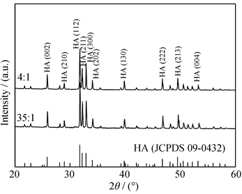
Fig. 1 XRD patterns of HA4 spherules and HA35 spherules
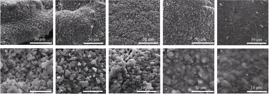
Fig. 2 SEM images of surface HA spheres with different initial ratios of Chitin/HAa1, b1: HA4; a2, b2: HA10; a3, b3: HA20; a4, b4: HA30; a5, b5: HA35
| HA/Chitin | Micro-porosity/% | BET/(m2·g-1) |
|---|---|---|
| 4/1 | 35±0.8 | 152.49 |
| 10/1 | 30.1±0.9 | / |
| 20/1 | 25.7±0.4 | 86.39 |
| 30/1 | 19.6±0.5 | / |
| 35/1 | 10.4±0.7 | 17.25 |
Table 1 Microporosity and specific surface area of HA spheres with different ratios of HA/Chitin
| HA/Chitin | Micro-porosity/% | BET/(m2·g-1) |
|---|---|---|
| 4/1 | 35±0.8 | 152.49 |
| 10/1 | 30.1±0.9 | / |
| 20/1 | 25.7±0.4 | 86.39 |
| 30/1 | 19.6±0.5 | / |
| 35/1 | 10.4±0.7 | 17.25 |
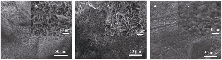
Fig. 3 SEM images of the surface of HA spheres (a, b) mineralized for 3 da1, b1: HA4; a2, b2: HA20; a3, b3: HA35
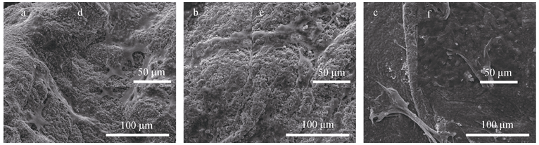
Fig. 4 SEM images of BM-MSCs cultured on HA spehres with different surface microstructures after 3 da, d: HA4; b, e: HA20; c, f: HA35
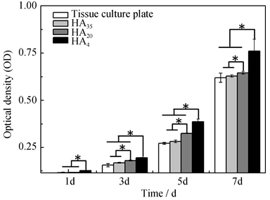
Fig. 5 Proliferation of BM-MSCs cultured on tissue culture plates and HA spheres with different surface microstructures at 1, 3, 5 and 7 d(* represents statistical difference, P<0.05)
| CD29 | CD44 | CD90 | |
|---|---|---|---|
| Control | 99.2% | 93% | 98.9% |
| HA4 | 89.4% | 67% | 90.9% |
| HA20 | 74.6% | 56% | 76.0% |
| HA35 | 66.9% | 40% | 59.7% |
Table 2 Surface characteristic markers expression of BM- MSCs cultured on HA spheres with different surface microstructures after 7 d
| CD29 | CD44 | CD90 | |
|---|---|---|---|
| Control | 99.2% | 93% | 98.9% |
| HA4 | 89.4% | 67% | 90.9% |
| HA20 | 74.6% | 56% | 76.0% |
| HA35 | 66.9% | 40% | 59.7% |

Fig. 6 Expression of ALP of BM-MSCs cultured on HA spheres with different surface microstructures at 7 d(* represents statistical difference, P<0.05)
| [1] | ROY D M, LINNEHAN S K.Hydroxyapatite formed from coral skeletal carbonate by hydrothermal exchange.Nature, 1974, 247(438): 220-222. |
| [2] | LI Y, TJANDRA W, TAM K C.Synthesis and characterization of nanoporous hydroxyapatite using cation surfactants as templates.Mater. Res. Bull. , 2008, 43(8/9): 2318-2326. |
| [3] | YANG Z, YUAN H, TONG W, et al.Osteogenesis in extraskeletally implanted porous calcium phosphate ceramics: variability among different kinds of animals.Biomaterials, 1996, 17(22): 2131-2137. |
| [4] | ZHI W, ZHANG C, DUAN K, et al.A novel porous bioceramics scaffold by accumulating hydroxyapatite spherulites for large bone tissue engineering in vivo.Ⅱ. Construct large volume of bone grafts.J. Biomed. Mater. Res., 2014, 108(8): 2491-2501. |
| [5] | TRIPATHI G, BASU B.A porous hydroxyapatite scaffold for bone tissue engineering: Physico-mechanical and biological evaluations.Ceramics International, 2012, 38(1): 341-349. |
| [6] | WANG H, ZHI W, LU X, et al.Comparative studies on ectopic bone formation in porous hydroxyapatite scaffolds with complementary pore structures.Acta Biomaterialia, 2013, 9(9): 8413-8421. |
| [7] | TEIXEIRA S, RODRIGUEZ M A, PENA P, et al.Physical characterization of hydroxyapatite porous scaffolds for tissue engineering.Materials Science and Engineering: C, 2009, 29(5): 1510-1514. |
| [8] | BARRADAS A, YUAN H, BLITTERSWIJK C A, et al.Osteoinductive biomaterials: current knowledge of properties, experimental models and biological mechanisms.European Cells and Materials, 2011, 21: 407-429. |
| [9] | ZHOU H, WU T, DONG X, et al.Adsorption mechanism of BMP-7 on hydroxyapatite (001) surface.Biochem. Biophys. Res. Commun., 2007, 361(1): 91-96. |
| [10] | WOODARD J R, HILLDORE A J, LAN S K, et al.The mechanical properties and osteoconductivity of hydroxyapatite bone scaffolds with multi-scale porosity.Biomaterials, 2007, 28(1): 45-54. |
| [11] | HABIBOVIC P, YUAN H, VAN DER VALK C M, et al. 3D microenvironment as essential element for osteoinduction by biomaterials.Biomaterials, 2005, 26(17): 3565-3575. |
| [12] | YAMASAKI H, SAKAI H.Osteogenic response to porous hydroxyapatite ceramics under the skin of dogs.Biomaterials, 1992, 13(5): 308-312. |
| [13] | ZHANG J, LUO X, BARBIERI D, et al.The size of surface microstructures as an osteogenic factor in calcium phosphate ceramics.Acta Biomaterialia, 2014, 10(7): 3254-3263. |
| [14] | PONADER S, VAIRAKTARIS E, HEINL P, et al.Effects of topographical surface modifications of electron beam melted Ti-6Al-4V titanium on human fetal osteoblasts. J. Biomed. Mater. Res. Part A, 2008, 84A(4): 1111-1119. |
| [15] | NATH S, TRIPATHI R, BASU B.Understanding phase stability, microstructure development and biocompatibility in calcium phosphate-titania composites, synthesized from hydroxyapatite and titanium powder mix.Mater. Sci. Eng. C, 2009, 29(1): 97-107. |
| [16] | ZHOU H, LEE J.Nanoscale hydroxyapatite particles for bone tissue engineering.Acta Biomater., 2011, 7(7): 2769-2781. |
| [17] | LI D, LU X, LIN H, et al.Chitosan/bovine serum albumin co- micropatterns on functionalized titanium surfaces and their effects on osteoblasts. J.Mater. Sci. Mater. Med., 2013, 24(2): 489-502. |
| [18] | ZHANG J, BARBIERI D, TEN HOOPEN H, et al.Microporous calcium phosphate ceramics driving osteogenesis through surface architecture.Journal of Biomedical Materials Research Part A. 2015, 103(3): 1188-1199. |
| [19] | 翁杰, 彭谦, 屈树新, 等. 一种制备多孔结构球形颗粒无机材料制品的方法. 中国发明专利, ZL200410081377.7. 2006. |
| [20] | PENG Q, JIANG F, HUANG P, et al.A novel porous bioceramics scaffold by accumulating hydroxyapatite spherulites for large bone tissue engineering in vivo. I: preparation and characterization of scaffold. J.Biomed. Mater. Res., 2010, 93(3): 920-929. |
| [21] | GUO LAI-YANG, ZHANG JING-WEI, ZHAO JING, et al.Preparation and characterization of porous scaffolds with favourable interpore connecting.Journal of Inorganic Materials, 2011, 26(1): 17-21. |
| [22] | YUAN H, FERNANDES H, HABIBOVIC P, et al.Osteoinductive ceramics as a synthetic alternative to autologous bone grafting.Proc. Natl. Acad. Sci. U S A. 2010, 107(31): 13614-13619. |
| [23] | GALLI C, PIEMONTESE M, LUMETTI S, et al.The importance of WNT pathways for bone metabolism and their regulation by implant topography.European Cells and Materials. 2012, 24: 46-59. |
| [24] | HABIBOVIC P, SEES T M, VAN DEN DOEL M A, et al. Osteoinduction by biomaterials-physicochemical and structural influences.J. Biomed. Mater. Res. A, 2006, 77(4): 747-762. |
| [25] | NAKAYAMA G R, CATON M C, NOVA M P, et al.Assessment of the Alamar Blue assay for cellular growth and viability in vitro. J.Immunol. Methods, 1997, 204(2): 205-208. |
| [26] | CHOU L, FIRTH J D, UITTO V J, et al.Substratum surface topography alters cell shape and regulates fibronectin mRNA level, mRNA stability, secretion and assembly in human fibroblasts.Journal of Cell Science. 1995, 108(4): 1563-1573. |
| [27] | PERIZZOLO D, LACEFIELD W R, BRUNETTE D M.Interaction between topography and coating in the formation of bone nodules in culture for hydroxyapatite-and titanium-coated micromachined surfaces.Journal of Biomedical Materials Research, 2001, 56(4): 494-503. |
| [28] | HARTING M T, JIMENEZ F, PATI S, et al.Immunophenotype characterization of rat mesenchymal stromal cells.Cytotherapy, 2008, 10(3): 243-253. |
| [29] | OH S, BRAMMER K S, LI Y S J, et al. Stem cell fate dictated solely by altered nanotube dimension.Proc. Natl. Acad. Sci. U S A, 2009, 106(7): 2130-2135. |
| [1] | AN Ran, LIN Si, GUO Shigang, ZHANG Chong, ZHU Shun, HAN Yingchao. Iron-doped Nano-hydroxyapatite: Preparation and Ultraviolet Absorption Performance [J]. Journal of Inorganic Materials, 2025, 40(5): 457-465. |
| [2] | LI Zhenghao, LI Jingming, ZHANG Yuxiang, YUAN Bo, ZHANG Kai, ZHU Xiangdong. Preparation and in vitro Osteogenic Activity Evaluation of Mn/nHA Coated CF/PEEK Composite [J]. Journal of Inorganic Materials, 2025, 40(10): 1145-1152. |
| [3] | LI Chengyu, DING Ziyou, HAN Yingchao. In vitro Antibacterial and Osteogenic Properties of Manganese Doped Nano Hydroxyapatite [J]. Journal of Inorganic Materials, 2024, 39(3): 313-320. |
| [4] | LIU Yan, ZHANG Yufan, WANG Ximan, LI Ting, MA Wenting, YANG Fuwei, CHEN Liang, ZHAO Dongyue, YAN Xiaoqin. Consolidation of Fragile Weathered Bone Relics Using Hydroxyapatite Material as Consolidant [J]. Journal of Inorganic Materials, 2023, 38(11): 1345-1354. |
| [5] | CHEN Yaling, SHU Song, WANG Shaoxin, LI Jianjun. Mn-HAP SCR Catalyst: Preparation and Sulfur Resistance [J]. Journal of Inorganic Materials, 2022, 37(10): 1065-1072. |
| [6] | ZHU Yutong, TAN Peijie, LIN Hai, ZHU Xiangdong, ZHANG Xingdong. Injectable Hyaluronan/Hydroxyapatite Composite: Preparation, Physicochemical Property and Biocompatibility [J]. Journal of Inorganic Materials, 2021, 36(9): 981-990. |
| [7] | LIN Ziyang, CHANG Yuchen, WU Zhangfan, BAO Rong, LIN Wenqing, WANG Deping. Different Simulated Body Fluid on Mineralization of Borosilicate Bioactive Glass-based Bone Cement [J]. Journal of Inorganic Materials, 2021, 36(7): 745-752. |
| [8] | WU Zhongcao, HUAN Zhiguang, ZHU Yufang, WU Chengtie. 3D Printing and Characterization of Microsphere Hydroxyapatite Scaffolds [J]. Journal of Inorganic Materials, 2021, 36(6): 601-607. |
| [9] | WU Yonghao, LI Xiangfeng, ZHU Xiangdong, ZHANG Xingdong. Construction of Hydroxyapatite Nanoceramics with High Mechanical Strength and Efficiency in Promoting the Spreading and Viability of Osteoblasts [J]. Journal of Inorganic Materials, 2021, 36(5): 552-560. |
| [10] | SONG Keke, HUANG Hao, LU Mengjie, YANG Anchun, WENG Jie, DUAN Ke. Hydrothermal Preparation and Characterization of Zn, Si, Mg, Fe Doped Hydroxyapatite [J]. Journal of Inorganic Materials, 2021, 36(10): 1091-1096. |
| [11] | SHAO Yueting, ZHU Yingjie, DONG Liying, CAI Anyong. Nanocomposite “Xuan Paper” Made from Ultralong Hydroxyapatite Nanowires and Cellulose Fibers and Its Anti-mildew Properties [J]. Journal of Inorganic Materials, 2021, 36(1): 107-112. |
| [12] | SUN Tuanwei,ZHU Yingjie. One-step Solvothermal Synthesis of Strontium-doped Ultralong Hydroxyapatite Nanowires [J]. Journal of Inorganic Materials, 2020, 35(6): 724-728. |
| [13] | LIU Ziyang, GENG Zhen, LI Zhaoyang. Preparing Biomedical CaCO3/HA Composite with Oyster Shell [J]. Journal of Inorganic Materials, 2020, 35(5): 601-607. |
| [14] | DAI Zhao,WANG Ming,WANG Shuang,LI Jing,CHEN Xiang,WANG Da-Lin,ZHU Ying-Chun. Zirconia Reinforced Trace Element Co-doped Hydroxyapatite Coating [J]. Journal of Inorganic Materials, 2020, 35(2): 179-186. |
| [15] | FU Ya-Kang,WENG Jie,LIU Yao-Wen,ZHANG Ke-Hong. hBMP-2 Contained Composite Coatings on Titanium Mesh Surface: Preparation and hBMP-2 Release [J]. Journal of Inorganic Materials, 2020, 35(2): 173-178. |
| Viewed | ||||||
|
Full text |
|
|||||
|
Abstract |
|
|||||