Journal of Inorganic Materials ›› 2015, Vol. 30 ›› Issue (1): 53-58.DOI: 10.15541/jim20140178
• Orginal Article • Previous Articles Next Articles
WANG Jun1, ZHANG Bao-Lin1, YANG Gao1, WANG Lei2, XIE Song-Bo1, LI Xuan1, GAO Fa-Bao2
Received:2014-04-08
Revised:2014-06-20
Published:2015-01-20
Online:2014-12-29
About author:WANG Jun. E-mail: wjs19892008@126.com
Supported by:CLC Number:
WANG Jun, ZHANG Bao-Lin, YANG Gao, WANG Lei, XIE Song-Bo, LI Xuan, GAO Fa-Bao. Synthesis and Application of Magnetic Iron Oxide Nanoparticles as High Efficiency Magnetic Resonance Imaging Contrast Agent[J]. Journal of Inorganic Materials, 2015, 30(1): 53-58.

Fig. 1 XRD pattern of iron oxide nanoparticles prepared at 260℃ coated with PEG

Fig. 2 TEM image and size distribution of PEG-SPIONs prepared at 260℃
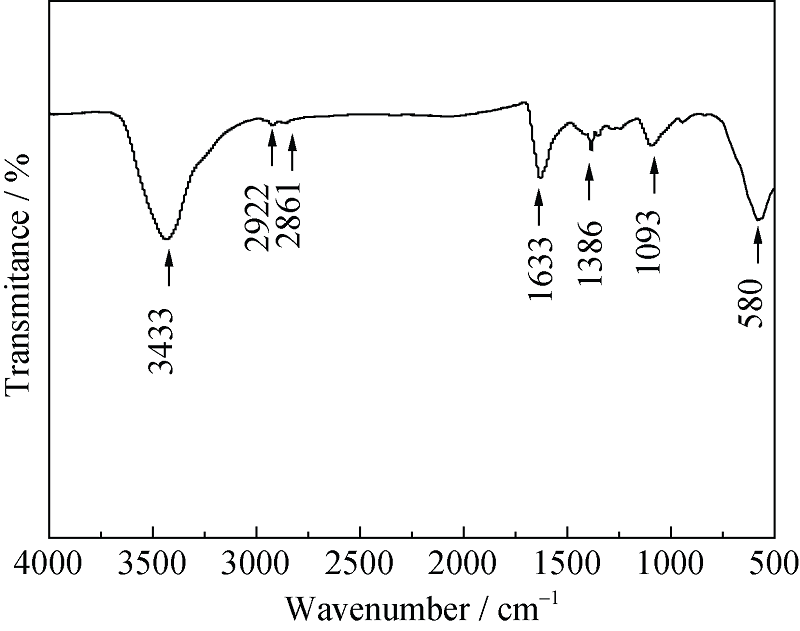
Fig. 3 FTIR spectrum of PEG-SPIONs prepared at 260℃

Fig. 4 Hydrodynamic size(a) and Zeta potential(b) of the iron oxide nanoparticles coated with PEG prepared at 260℃ in deionized water

Fig. 5 M-H curve of PEG-SPIONs at 300 K
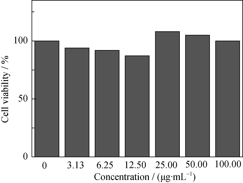
Fig. 6 Cell viabilities of MCF-7 cells after incubation with different concentrations of PEG-SPIONs for 24 h

Fig. 7 T2 weight MR images (a) and the T2 relaxiation rate (b) of PEG-SPIONs with different Fe concentrations
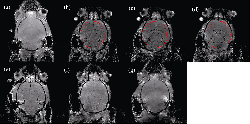
Fig. 8 T2* MR images of the mouse brain before and after intravenous injection of PEG-SPIONs. (a) before injection, (b) 5 min, (c) 10 min, (d) 30 min, (e) 1 h, (f) 4 h and (g) 24 h after injection
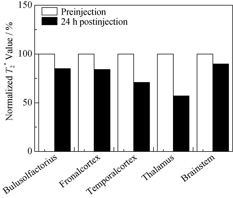
Fig. 9 Relative T2* value of different brain regions extracted from T2* MR images of mouse brains before injection and 24 h after injection PEG-SPIONs
| [1] | LAMKOWSKY M C, GEPPERT M, SCHMIDT M M, et al.Magnetic field-induced acceleration of the accumulation of magnetic iron oxide nanoparticles by cultured brain astrocytes.J. Biomed. Mater. Res., 2012, 100(2): 323-334. |
| [2] | YANG X Q, HONG H, GRAILER J J, et al.cRGD-functionalized, DOX-conjugated, and 64 Cu-labeled superparamagnetic iron oxide nanoparticles for targeted anticancer drug delivery and PET/MR imaging. Biomaterials, 2011, 32(17): 4151-4160. |
| [3] | XU H, CHENG L, WANG C, et al.Polymer encapsulated upconversion nanoparticle/iron oxide nanocomposites for multimodal imaging and magnetic targeted drug delivery.Biomaterials, 2011, 32(35): 9364-9373. |
| [4] | LEE N, HYEON T.Designed synthesis of uniformly sized iron oxide nanoparticles for efficient magnetic resonance imaging contrast agents.Chem. Soc. Rev., 2012, 41(7): 2575-2589. |
| [5] | CASULA M F, FLORIS P, INNOCENTI C, et al.Magnetic resonance imaging contrast agents based on iron oxide superparamagnetic ferrofluids.Chem. Mater., 2010, 22(5): 1739-1748. |
| [6] | SUN S, ZENG H, ROBINSON D B, et al.Monodisperse MFe2O4 (M = Fe, Co, Mn) nanoparticles.J. Am. Chem. Soc., 2004, 126(1): 273-279. |
| [7] | KIM B H, LEE N, KIM H, et al.Large-scale synthesis of uniform and extremely small-sized iron oxide nanoparticles for high-resolution T1 magnetic resonance imaging contrast agents.J. Am. Chem. Soc., 2011, 133(32): 12624-12631. |
| [8] | NA H B, LEE I S, SEO H, et al.Versatile PEG-derivatized phosphine oxide ligands for water-dispersible metal oxide nanocrystals.Chem. Commun., 2007, 48: 5167-5169. |
| [9] | XU F, ZHANG X, XIE Y, et al.Morphology control of γ-Fe2O3 nanocrystals via PEG polymer and accounts of its Mössbauer study.J. Colloid. Interface. Sci., 2003, 260(1): 160-165. |
| [10] | MUTHIAH M, PARK I K, CHO C S, Surface modification of iron oxide nanoparticles by biocompatible polymers for tissue imaging and targeting.Biotechnol. Adv., 2013, 31(8): 1224-1236. |
| [11] | TU Z J, ZHANG B L, YANG G, et al.Synthesis of poly (ethylene glycol) and poly (vinyl pyrrolidone) co-coated superparamagnetic iron oxide nanoparticle as a pH-sensitive release drug carrier.Colloid. Surface. A, 2013, 436: 854-861. |
| [12] | MINATI L, MICHELI V, ROSSI B, et al.Application of factor analysis to XPS valence band of superparamagnetic iron oxide nanoparticles.Appl. Surf. Sci., 2011, 257(24): 10863-10868. |
| [13] | PARK J, AN K,.HWANG Y, et al.Ultra-large-scale syntheses of monodisperse nanocrystals.Nat. Mater., 2004, 3(12): 891-895. |
| [14] | GONCALVES R H, CARDOSO C A, LEITE E R.Synthesis of colloidal magnetite nanocrystals using high molecular weight solvent.J. Mater. Chem., 2010, 20(6): 1167-1172. |
| [15] | LIU Q X, XU Z H.Self-assembled monolayer coatings on nanosized magnetic particles using 16-mercaptohexadecanoic acid. Langmuir, 1995, 11(12): 4617-4622. |
| [16] | XU Y, QIN Y, Palchoudhury S, et al.Water-soluble iron oxide nanoparticles with high stability and selective surface functionality.Langmuir, 2011, 27(14): 8990-8997. |
| [17] | XUE HONG-TAO, SHEN SHUI-FA, PAN HAI-BO, et al.Preparation and characterization of mesoporous iron oxide. J. Inorg. Mater., 2009, 24(3):576-580. |
| [18] | MAITY D, KALE S N, KAUL-GHANEKAR R, et al.Studies of magnetite nanoparticles synthesized by thermal decomposition of iron (III) acetylacetonate in tri (ethylene glycol). J. Magn. Magn. Mater., 2009, 321(19): 3093-3098. |
| [19] | MONDINI S, CENEDESE S, MARINONI G, et al.One-step synthesis and functionalization of hydroxyl-decorated magnetite nanoparticles.J. Colloid. Interface. Sci., 2008, 322(1): 173-179. |
| [20] | GUN S, EDIRISINGHE M, STRIDE E, et al.Encapsulation of superparamagnetic iron oxide nanoparticles in poly- (lactide- coglycolic acid) microspheres for biomedical applications.Mater. Sci. Eng. C, 2013, 33(6): 3129-3137. |
| [21] | QIAO R R, YANG C H, GAO M Y.Superparamagnetic iron oxide nanoparticles: from preparations to in vivo MRI applications. J. Mater. Chem., 2009, 19(35): 6274-6293. |
| [22] | ZENG Q, BAKER I, HOOPES J.The heating effects of dextran coated iron oxides.Mater. Res. Soc. Symp. Proc., 2007, 962(20): 10-16. |
| [23] | ZHANG B L, TU Z J, ZHAO F Y, et al.Superparamagnetic iron oxide nanoparticles prepared by using an improved polyol method.Appl. Surf. Sci., 2013, 266: 375-379. |
| [24] | JUN Y, SEO J, CHEON J.Nanoscaling laws of magnetic nanoparticles and their applicabilities in biomedical sciences.Acc. Chem. Res., 2008, 41(2): 179-189. |
| [25] | ZHAO F Y, ZHANG B L, FENG L Y.Preparation and magnetic properties of magnetite nanoparticles.Mater. Lett., 2012, 68: 112-114. |
| [26] | Hu F Q, MACRENARIS K W, WATERS E A, et al.water-soluble magnetite nanoparticles with high relaxivity for magnetic resonance imaging,J. Phys. Chem. C, 2009, 113(49): 20850-20855. |
| [27] | TROMSDORF U I, BRUNS O T, SALMEN S C, et al.A highly effective, nontoxic T1 MR contrast agent based on ultrasmall PEGylated iron oxide nanoparticles.Nano Lett., 2009, 9(12): 4434-4440. |
| [28] | LEE N, HYEON T.Designed synthesis of uniformly sized iron oxide nanoparticles for efficient magnetic resonance imaging contrast agents. Chem. Soc. Rev., 2012, 41(7): 2575-2589. |
| [1] | YU Man, GAO Rongyao, QIN Yujun, AI Xicheng. Influence of Upconversion Luminescent Nanoparticles on Hysteresis Effect and Ion Migration Kinetics in Perovskite Solar Cells [J]. Journal of Inorganic Materials, 2024, 39(4): 359-366. |
| [2] | WANG Tongyu, RAN Haofeng, ZHOU Guangdong. Defect-induced Analogue Resistive Switching Behavior in FeOx-based Memristor and Synaptic Paired-pulse Facilitation Feature [J]. Journal of Inorganic Materials, 2023, 38(4): 437-444. |
| [3] | YU Yefan, XU Ling, NI Zhongbing, SHI Dongjian, CHEN Mingqing. Prussian Blue Modified Biochar: Preparation and Adsorption of Ammonia Nitrogen from Sewage [J]. Journal of Inorganic Materials, 2023, 38(2): 205-212. |
| [4] | WANG Xiaojun, XU Wen, LIU Runlu, PAN Hui, ZHU Shenmin. Preparation and Properties of Ag@C3N4 Photocatalyst Supported by Hydrogel [J]. Journal of Inorganic Materials, 2022, 37(7): 731-740. |
| [5] | MA Hui, TAO Jianghui, WANG Yanni, HAN Yu, WANG Yabin, DING Xiuping. Gold Nanoparticles Supported on Silica & Titania Hybrid Mesoporous Spheres and Their Catalytic Performance Regulation [J]. Journal of Inorganic Materials, 2022, 37(4): 404-412. |
| [6] | SU Li, YANG Jianping, LAN Yue, WANG Lianjun, JIANG Wan. Interface Design of Iron Nanoparticles for Environmental Remediation [J]. Journal of Inorganic Materials, 2021, 36(6): 561-569. |
| [7] | ZHANG Yiqing,ZHANG Shujuan,WAN Zhengrui,MO Han,WANG Niangui,ZHOU Liqun. RuFe Nanoparticles Modified Sheet-like BiVO4 : High-efficient Synergistic Catalyst for Ammonia Borane Hydrolytic Dehydrogenation [J]. Journal of Inorganic Materials, 2020, 35(7): 809-816. |
| [8] | SHAO Kang,WANG Tao,WEN Yingting,TENG Yuanjie,LIU Huijun,PAN Zaifa. Modulation of Morphology and Luminescence Property of NaBiF4:Yb3+/Er3+ Upconversion Nanoparticles by Organic Ligands [J]. Journal of Inorganic Materials, 2020, 35(4): 447-453. |
| [9] | GUO Si-Lin, KANG Shuai, LU Wen-Qiang. Ge Nanoparticles in MXene Sheets: One-step Synthesis and Highly Improved Electrochemical Property in Lithium-ion Batteries [J]. Journal of Inorganic Materials, 2020, 35(1): 105-111. |
| [10] | LI Sheng-Song, ZHENG Yong-Chao, MENG Shu-Lin, WU Li-Zhu, ZHONG Jin- Yi, ZHAO Chong-Lin. Core/Shell Quantum Dots and Au Nanoparticles Assembly for Effective Detection of Nerve Agent Mimic [J]. Journal of Inorganic Materials, 2019, 34(8): 893-898. |
| [11] | ZHU Meng-Meng, LI Guo-Hua, ZHANG Xue-Ming, ZHAI Jia-Xin, GAN Si-Ping, SONG Xiao. Boron Nitride Nanosheets Supported Cu2O Nanoparticles: Synthesis and Catalytic Reduction for 4-nitrophenol [J]. Journal of Inorganic Materials, 2019, 34(8): 817-826. |
| [12] | CHENG Tian-Sheng, PAN Jiong, XU Ying-Ying, BAO Qun-Qun, HU Ping, SHI Jian-Lin. Synthesis of Zn, Mn doped Fe3O4 Nanoparticles with Tunable Size [J]. Journal of Inorganic Materials, 2019, 34(8): 899-903. |
| [13] | WANG Ya, SUO Hong-Li, LIU Min, WANG Tian-Tian, KAUSAR Shaheen, XU Yan, MA Lin. Property of YBCO Films Doping with Positive and Negative Lattice Mismatch Nanoparticles [J]. Journal of Inorganic Materials, 2019, 34(8): 857-861. |
| [14] | WANG Ya, SUO Hong-Li, MAO Lei, LIU Min, MA Lin, WANG Yi, KAUSAR Shaheen, ZHOU Yu-Qi. Flux Pinning Mechanism of Nb-doped YBCO Film [J]. Journal of Inorganic Materials, 2019, 34(10): 1055-1059. |
| [15] | LIU Huan-Long, ZHAO Wei, LI Rui-Zhe, HUANG Xie-Yi, TANG Yu-Feng, LI Dong-Mei, HUANG Fu-Qiang. Facile Synthesis of Reduced Graphene Oxide In-situ Wrapped MnTiO3 Nanoparticles for Excellent Lithium Storage [J]. Journal of Inorganic Materials, 2018, 33(9): 1022-1028. |
| Viewed | ||||||
|
Full text |
|
|||||
|
Abstract |
|
|||||