

相比之下, 无机保护材料具有良好耐候性和兼容性, 更适合于加固保护骨质文物[17-18], 其研究与应用也逐渐兴起。葛丹阳等[19]探索利用微生物沉积碳酸钙加固考古骨的方法, 发现巴士芽孢杆菌能在考古骨表面生成碳酸钙, 弥合其表面裂隙。Palazzo 等[20]研究发现纳米氢氧化钙的丙醇分散液可渗入风化骨片中的微小孔隙, 并起到填充加固作用。杨富巍等[21]则将纳米氢氧化钙的醇悬浮分散液和磷酸铵溶液先后渗入骨质文物, 利用两者间的化学反应在其内部生成具有加固作用的磷灰石连续相。如前所述, 磷灰石本身就是骨质文物的主要化学成分, 从材料兼容性角度, 这种“以骨补骨”的方法更加安全有效[22]。但是, 磷酸铵溶液与氢氧化钙反应时会释放对操作人员有害的刺激性氨气, 而且将钙和磷分别导入的方法会导致两者在骨质文物内部分布不均匀, 造成加固效果不稳定。
为解决上述问题, 本研究按一定计量比混合亚微米氧化钙-磷酸氢钙制备成乙醇悬浮分散液, 以此作为骨质文物加固保护材料, 取得了优良的加固效果。
1 实验方法
1.1 试剂与材料
二水磷酸氢钙(CaHPO4·2H2O, AR), 氧化钙(CaO, AR), 无水乙醇(CH3CH2OH, AR)均购自国药集团化学试剂有限公司。用于制作模拟风化脆弱骨样品的猪新鲜肩胛骨为市售。发掘出土的风化脆弱骨片来自西北大学古人类骨骼标本库。
1.2 氧化钙-磷酸氢钙乙醇悬浮分散液制备
图1
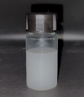
图1
25%悬浮浓度的氧化钙-磷酸氢钙乙醇分散液
Fig. 1
Calcium oxide-calcium hydrogen phosphate dispersion in alcohol at concentration of 25%
1.3 模拟风化骨样品制备
1.4 模拟风化骨样品加固
将氧化钙-磷酸氢钙的乙醇分散液分别以滴渗、涂刷、浸泡的方式引入脆弱骨样品内部。然后, 使用蒸馏水对脆弱骨样品进行二次渗透处理, 使氧化钙、磷酸氢钙发生反应生成羟基磷灰石连续相而达到加固的目的。
1.5 实验步骤
首先, 按照表1配比制备质量分数为25%的氧化钙-磷酸氢钙乙醇悬浮分散液备用。
表1 实验分组方案
Table 1
| Group | Mass ratio | |||||
|---|---|---|---|---|---|---|
| 1 : 1 | 1 : 3 | 1 : 4 | 1 : 5 | 1 : 6 | 1 : 7 | |
| Brushing | 1 | 2 | 3 | 4 | 5 | 6 |
| Soaking | 7 | 8 | 9 | 10 | 11 | 12 |
| Dripping | 13 | 14 | 15 | 16 | 17 | 18 |
Note: The blank control group was labeled as 19
然后, 利用下述三种方式施加悬浮分散液。
(1)涂刷 利用软毛刷将悬浮液反复涂刷于脆弱骨样品表面, 直至脆弱骨样品不再吸收悬浮液。
(2)浸泡 将脆弱骨样品置于悬浮液中浸渗处理, 直至脆弱骨样品不再吸收悬浮液。
(3)滴渗 利用胶头滴管吸取悬浮液, 将其滴 加于脆弱骨样品表面, 直至脆弱骨样品不再吸收悬浮液。
最后, 将蒸馏水滴加于脆弱骨样品表面直至其不再吸收, 并在25.0 ℃、60.0%湿度环境中静置1 d。
1.6 加固效果表征方法
采用分析天平(FA2204C, 上海天美天平仪器有限公司, 精度0.0001 g)测得样品质量变化。分别称取脆弱骨样品加固后的质量(记为m1)和加固前脆弱骨样品质量(记为m0), 使用以下公式计算质量增长率ΔM。
采用精密色度仪(WSC-2B, 上海电物理光学仪器有限公司)测得样品色差, 脆弱骨样品加固前后外观的改变采用色差变化表示:
其中, ΔE为色差, L为亮度值, a为黄绿值, b为黄蓝值。利用孔隙率测试仪(MZ-C300, 秒准科技(深圳)有限公司)测得样品开放孔隙率(PO)、密度(SG);通过样品断面的胶带试验(STT)测定加固后样品的强度, 加固强度(Strength, Str)按下式计算:
式中, m0、m1分别代表样品初始质量和胶带从样品断面上黏接下颗粒或碎块后的质量, S代表胶带的面积。利用扫描电子显微镜(TESCAN MIRA3, 捷克TESCAN公司, 加速电压10.00 kV, 工作距离7.6~11.9 mm)测定样品微观结构和元素组成;利用X射线衍射仪分析样品物相组成 (XRD, SmartLAB, 日本株式会社理学公司, 管电压为40 kV, 管电流为150 mA, 扫描范围为5°~90°, 步长0.01°, 扫描速率为10 (°)·min-1)。采用便携式超景深显微镜观察样品截面(4800-b, 深圳市宗源伟业科技有限公司)。
2 结果与分析
2.1 加固前后脆弱骨样品外观变化
图2
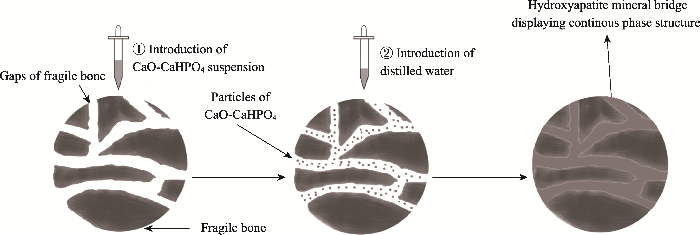
图2
基于氧化钙-磷酸氢钙乙醇分散液的风化脆弱骨样品加固机理示意图
Fig. 2
Consolidation mechanism of weathered bones using calcium oxide-calcium hydrogen phosphate dispersion in alcohol as consolidant
图3
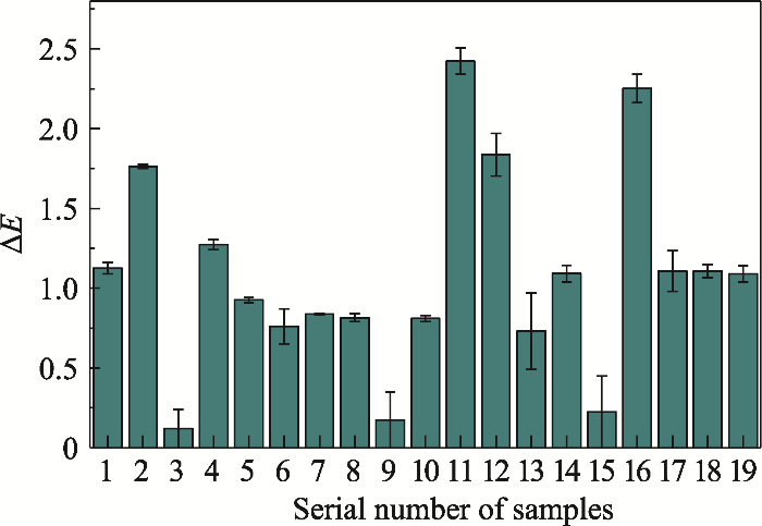
图3
脆弱骨样品加固处理前后色差
Fig. 3
Color difference of fragile bone samples after consolidation treatment
Samples 1 to 19 are introduced in
2.2 加固前后骨样品质量增长率
加固剂在脆弱骨样品内部的留存量可用质量增长率表示。质量增长率越大, 证明加固材料留存于脆弱骨样品中的量越大, 加固效果越好;反之则加固效果越弱。
图4
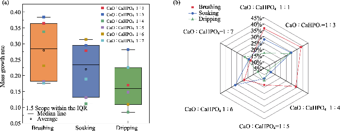
图4
加固方式和加固剂配比对脆弱骨样品质量增长率的影响
Fig. 4
Effects of consolidation way and consolidant ratio on the bone weight increment
(The rectangle is Interquartile Range (IQR), which is used to mark data outliers)
(a) Consolidation way; (b) Consolidant ratio
不同加固方式造成加固剂渗入量的差异应该是多种因素综合作用的结果。涂刷方式下, 毛细吸收和重力是加固剂进入脆弱骨样品内部的双重动力, 而且刷涂方式下加固剂在脆弱骨表面分布相对均匀, 不会因局部集中而阻塞孔隙, 影响后续渗透。浸泡方式下加固剂仅靠毛细吸收作用进入脆弱骨样品内部, 效果不如双重动力促进渗透的刷涂法。而滴渗方式下, 加固剂渗入脆弱骨样品虽然也是毛细吸收和重力双重动力, 但难以避免加固剂局部集中而快速阻塞孔隙, 影响加固剂的后续渗入, 致使其效果不如浸泡法。
2.3 加固前后孔隙率和密度变化
脆弱骨样品孔隙率采用水浸法测试。由图5(a)可知, 加固剂中氧化钙和磷酸氢钙的配比为1 : 3时, 三种施加方式均能减小脆弱骨样品的开放孔隙率, 并以刷涂方式最为显著。这与质量变化分析中样品质量增长的规律一致, 说明孔隙率降低是加固剂进入脆弱骨内部的直接结果。
图5
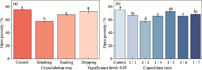
图5
加固方式和加固剂配比对脆弱骨样品孔隙率的影响
Fig. 5
Effects of consolidation way and consolidant ratio on the bone porosity
(a) Consolidation way; (b) Consolidant ratio
图6
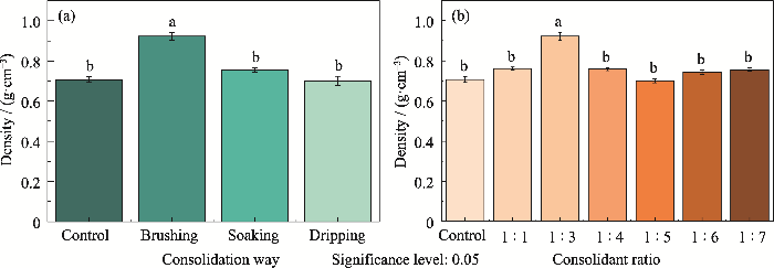
图6
加固处理方式和加固剂配比对脆弱骨样品密度的影响
Fig. 6
Effects of consolidation way and consolidant ratio on the bone density
(a) Consolidation way; (b) Consolidant ratio
图7
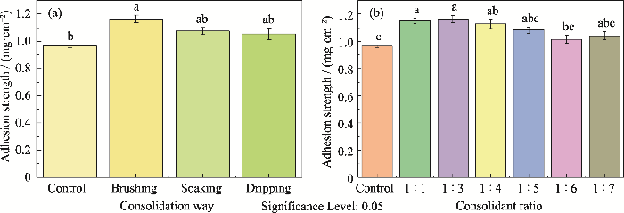
图7
加固处理方式和加固剂配比对脆弱骨样品表面强度的影响
Fig. 7
Effects of consolidation method and consolidant ratio on surface strength of the bones
(a) Consolidation way; (b) Consolidant ratio
2.4 强度分析
从公式(3)可知, (m1-m0)/m0越小, 即胶带从脆弱骨样品断面上粘接下来的颗粒或碎块越少, 表明脆弱骨样品的内聚力越大, 加固强度越好。由图5可知, 加固剂1 : 3的配比和涂刷加固方式用于提高脆弱骨样品的表面内聚力和强度比较有利。该结果与前述质量及孔隙率变化结果一致。
2.5 形貌分析
图8
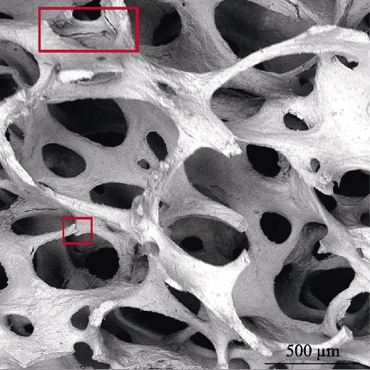
图8
加固前脆弱骨样品的显微形貌
Fig. 8
Morphology of fragile bone sample before consolidation treatment
The areas in red-framed indicate the fractures
图9
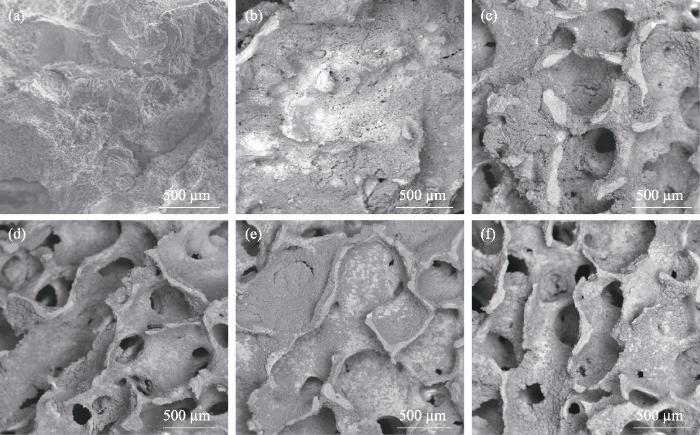
图9
加固处理后脆弱骨样品显微形貌
Fig. 9
Morphologies of fragile bone samples after consolidation treatments with mass ratios of calcium oxide to calcium hydrogen phosphate at 1 : 1 (a), 1 : 3 (b), 1 : 4 (c), 1 : 5 (d), 1 : 6 (e) and 1 : 7 (f), respectively
图10
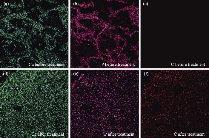
图10
加固处理前后脆弱骨样品EDS元素面分布结果
Fig. 10
EDS analysis results of fragile bone samples before and after consolidation treatment
(a, d) Ca; (b, e) P; (c, f) C, corresponding to
EDS元素分析如表2所示。加固前脆弱骨的主要元素为Ca、P、O和少量金属元素, 经加固后脆弱骨组成元素基本不变, 但是Ca、P的含量略有增加, 且新出现了C元素。它应该是加固剂中氧化钙水化并与空气中二氧化碳反应生成了碳酸钙所致。
表2 加固前后脆弱骨EDS元素分析结果
Table. 2
| Element | Weight ratio before consolidation/% | Weight ratio after consolidation/% |
|---|---|---|
| C | 0 | 5.62 |
| O | 56.93 | 56.32 |
| Na | 9.57 | 2.56 |
| Mg | 0.91 | 1.12 |
| P | 12.04 | 13.23 |
| Ca | 20.55 | 21.15 |
| Total | 100.00 | 100.00 |
图11
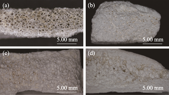
图11
加固处理前后脆弱骨样品的截面形貌
Fig. 11
Cross-sectional topographies of fragile bone samples before and after consolidation treatments
(a) Untreated; (b-d) Treated by brushing, soaking and dripping, respectively
图12
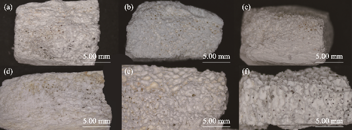
图12
不同配比的加固剂对脆弱骨加固处理后的样品截面形貌
Fig. 12
Cross-sectional topographies of fragile bone samples treated by different mass ratios of calcium oxide to calcium hydrogen phosphate
(a) 1 : 1; (b) 1 : 3; (c) 1 : 4; (d) 1 : 5; (e) 1 : 6; (f) 1 : 7
2.6 物相组成分析
图13
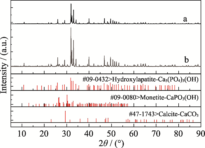
图13
脆弱骨样品加固处理前(a)后(b)的XRD图谱
Fig. 13
XRD results of bones before (a) and after (b) consolidation treatment
图14
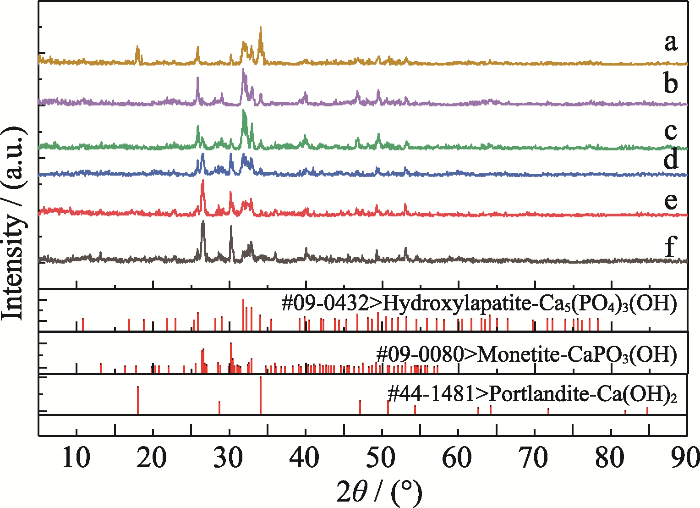
图14
加固剂质量配比对产物成分的影响
Fig. 14
Effect of consolidant ratio on final product
Ratios of calcium oxide to calcium hydrogen phosphate in line(a-f) are 1 : 1, 1 : 3, 1 : 4, 1 : 5, 1 : 6 and 1 : 7, respectively
表3 各配比下的物相比例
Table 3
| Ratio of CaO : CaHPO4 | Ca(OH)2 | CaHPO4 | Ca5(PO)4OH |
|---|---|---|---|
| 1 : 1 | +++ | - | ++ |
| 1 : 3 | - | - | ++++ |
| 1 : 4 | - | ++ | +++ |
| 1 : 5 | - | +++ | ++ |
| 1 : 6 | - | +++ | + |
| 1 : 7 | - | ++++ | + |
-: indicating less than 10%; +: indicating 10%-30%; ++: indicating 30%-50%; +++: indicating 50%-70%; ++++: indicating more than 70%
发掘出土风化脆弱骨片的加固效果见图15。加固处理前该骨片疏松多孔, 已经有酥粉脱落。采用1 : 3配比的氧化钙-磷酸氢钙悬浮液, 以表面刷涂方式加固处理后, 脆弱骨片中的孔隙得到有效填充, 脆弱骨片的整体强度也得到明显提高, 不再有酥粉脱落现象。
图15
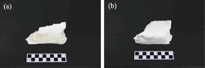
图15
考古发掘出土脆弱骨片加固处理前(a)后(b)的照片
Fig. 15
Photos of archaeological bone before (a) and after (b) consolidation treatment
2.7 讨论
研究采用氧化钙与磷酸氢钙混合物在乙醇中的悬浮分散液作为脆弱骨质文物的加固剂, 获得了良好的加固效果。设计这种保护方法基于以下三点:首先, 在乙醇中, 氧化钙和磷酸氢钙不会发生反应, 保证了其悬浮分散液的稳定性;其次, 乙醇的表面张力(0.0223 N/m)较低, 常温下还不到水(0.0727 N/m)的1/3, 使氧化钙和磷酸氢钙的混合物更容易渗入骨质文物内部;再次, 氧化钙和磷酸氢钙间反应本质上是离子反应, 蒸馏水是这类反应的良好介质, 可促进反应物电离出钙离子、磷酸根离子和氢氧根离子, 并生成羟基磷灰石连续相。该磷灰石连续相通过填充、弥合脆弱骨中的孔隙和裂缝, 使破碎的脆弱骨块重新结合而起到加固作用。另外, 羟基磷灰石中Ca/P摩尔比为1.67[38]。故以氧化钙和磷酸氢钙作为羟基磷灰石前驱物时, 两者的摩尔比应为2 : 3, 折合为质量比为1 : 3.5。本研究中氧化钙和磷酸氢钙的最佳配比为1 : 3, 说明此时氧化钙略微过量。这部分过量的氧化钙与水和空气中二氧化碳反应会进一步生成CaCO3, 起到协同加固的作用[39]。
本研究利用电镜、能谱、XRD、色差、质量、孔隙率、密度和断面强度等评估保护材料的填充性和加固效果, 均证实该材料具有较好的加固效果, 因而该方法具有较好的保护作用。人眼难以辨别加固处的色差, 加固材料能顺利而均匀地渗入脆弱骨质样品内部, 体现了加固材料良好的渗透性和填充性, 基本实现填充骨质样品内部孔隙的实验预期。氧化钙和磷酸氢钙的质量配比为1 : 3时, 目标产物的产率较高。经加固后的样品整体内聚力增大, 可有效抑制样品的酥粉掉渣现象, 达到了较为理想的加固效果。
3 结论
本研究探索了以氧化钙-磷酸氢钙的醇悬浮分散液为加固剂保护脆弱骨质文物的新方法, 发现:
1)当醇悬浮分散液中氧化钙和磷酸氢钙的质量配比为1 : 3、施加方式为表面刷涂时, 加固剂能较好地渗入脆弱骨样品内部;
2)加固剂中氧化钙和磷酸氢钙的反应产物羟基磷灰石能将脆弱骨样品内部的孔洞填充、黏接而起到加固保护作用。
3)利用该方法对脆弱骨进行加固处理后, 脆弱骨的孔隙率下降了17.3%, 质量、密度和表面黏结强度则分别提高了38.39%、34.49%和16.32%, 与空白组对照有显著差异, 且其色差也小于3.0, 符合文物保护要求。
参考文献
Methods for the assessment of human body composition: traditional and new
Characterisation of microbial attack on archaeological bone
Destruction of microstructure in archaeological bone: a case study from Portugal
The mechanical properties of artificially aged bone: probing the nature of the collagen-mineral bond
Durability of protective polymers: the effect of UV and thermal ageing
Plastic matters: an analytical procedure to evaluate the degradability of contemporary works of art

The most significant results concerning a chemical study to evaluate the degradability of polymeric components in four contemporary works of art, partially or completely realized in plastics, are presented and discussed in this paper. The procedure applied is mainly based on the use of Fourier transform IR and UV-vis spectroscopies and pyrolysis-gas chromatography/mass spectrometry, and consists of the following steps: (1) compositional analysis of the artworks, with particular attention to components which may have a negative effect on the overall ageing; (2) evaluation of the actual state of conservation; (3) investigation of the accelerated ageing of reference polymer samples; and (4) monitoring of the natural ageing of the artworks. On such a basis, the following could be concluded. Stage Evidence by Loris Cecchini is made of poly(ether urethane) elastomer which contains a high amount of phthalates. Their exudation gives a sticky appearance to the artwork and their removal during ageing is the main cause of the loss of flexibility. The latex used by Andrés Pinal for tailoring Traxe de Home is a natural polyisoprene, whose oxidative degradation accounts for the extensive deterioration and yellowing of the artwork. The plaster sculptures of 3D Bodyscans 1:9 by Karin Sander are coated with an aliphatic epoxy resin. Its oxidation with formation of amides is the cause of the surface yellowing. The adhesive used by Dario Villalba for Tierra, Ladrillo y Agua is a commercial poly(vinyl acetate). Simulated photoageing suggests a fast deterioration due to deacetylation and cross-linking, which possibly is the main reason for the actual detachment of debris from the support.
Smart cleaning of cultural heritage: a new challenge for soft nanoscience
The search for innovative, smart and performing cleaning agents is one of the main issues of modern conservation science. Nanosciences do not only provide solutions to this scientific field in terms of new materials but also change radically the approach to problems and challenges. In this feature article we review the most innovative nanostructured systems developed in the last decade for the cleaning of artworks together with some noteworthy case studies. Micelles, microemulsions, thickened complex fluids, and responsive gels that constitute the new "cleaning palette" for modern conservators are here presented and critically analyzed. The development of these smart nanostructured systems requires the comprehension of their behavior and interactions with other materials down to the nanoscale. In the last section of this manuscript we report on the most recent results from a study about the mechanism of polymer removal from porous artifacts using nanofluids, such as micelles or microemulsions. The rules of classical detergency do not fully address the polymer removal mechanism and a schematic model of the process is proposed.
New methodologies for the conservation of cultural heritage: micellar solutions, microemulsions, and hydroxide nanoparticles
Modern civilization's inherited artworks have a powerful impact on society, from political, sociological, and anthropological points of view, so the conservation of our Cultural Heritage is fundamental for conveying to future generations our culture, traditions, and ways of thinking and behaving. In the conservation of cultural artifacts, scientists intervene in the degradation of often unique handcrafts, resulting from a delicate balance of aging, unpredicted events, environmental conditions, and sometimes incorrect previous restoration treatments, the details of which are often not precisely known. Nanoscience and nanotechnology are revolutionizing materials science in a pervasive way, in a manner similar to polymer chemistry's revolution of materials science over the preceding century. The continuous development of novel nanoparticle-based materials and the study of physicochemical phenomena at the nanoscale are creating new approaches to conservation science, leading to new methodologies that can "revert" the degradation processes of the works of art, in most cases "restoring" them to their original magnificent appearance. Until recently, serendipity and experiment have been the most frequent design principles of formulations for either cleaning or consolidation of works of art. Accordingly, the past has witnessed a number of actively detrimental treatments, such as the application of acrylic and vinyl resins to wall paintings, which can irreversibly jeopardize the appearance (or even the continued existence) of irreplaceable works of art. Current research activity in conservation science is largely based on the paradigm that compatibility of materials is the most important prerequisite for obtaining excellent and durable results. The most advanced current methodologies are (i) the use of water-based micelles and microemulsions (neat or combined with gels) for the removal of accidental contaminants and polymers used in past restorations and (ii) the application of calcium hydroxide nanoparticles for the consolidation of works of art. In this Account, we highlight how conservation science can benefit from the conceptual and the methodological background derived from both soft (microemulsions and micelles for cleaning) and hard (nanoparticles for consolidation) nanoscience. A combination of different nanotechnologies allows today's conservators to provide, in each restoration step, interventions respectful of the physicochemical characteristics of the materials used by artists. The "palette" of methods provided by nanoscience is continuously enriching the field, and the development of novel nanomaterials and the study of nanoscale physicochemical phenomena will further improve the performance of restoration formulations and our comprehension of degradation mechanisms.
Comparative study between four consolidation systems suitable for archaeological bone artefacts
Indicators and ratings for the compatibility assessment of conservation actions
Ananalytical study of marble consolidation by oxalate precipitation using density, FTIR and powder-XRD measurements
Evaluation of the efficacy of traditional and nano paraloid b72 for pottery consolidation
In situ mechanical and molecular investigations of collagen/apatite biomimetic composites combining Raman spectroscopy and stress-strain analysis
Differential burning, recrystallization, and fragmentation of archaeological bone
A complementary approach using analytical pyrolysis to evaluate collagen degradation and mineral fossilisation in archaeological bones: the case study of Vicenne-Campochiaro necropolis (Italy)
Effect of the calcination temperature on the composition and microstructure of hydroxyapatite derived from human and animal bone
Conservation of bone relics using hydroxyapatite as protective material
