无机材料学报 ›› 2024, Vol. 39 ›› Issue (1): 90-98.DOI: 10.15541/jim20230151 CSTR: 32189.14.10.15541/jim20230151
所属专题: 【生物材料】抗菌与肿瘤治疗(202512)
何倩( ), 唐婉兰, 韩秉锟, 魏佳元, 吕文轩, 唐昭敏()
收稿日期:2023-03-23
修回日期:2023-04-24
出版日期:2024-01-20
网络出版日期:2023-09-12
通讯作者:
唐昭敏, 高级实验师. E-mail: tl8687@163.com作者简介:何 倩(1999-), 女, 硕士研究生. E-mail: 1084518887@qq.com
基金资助:
HE Qian(), TANG Wanlan, HAN Bingkun, WEI Jiayuan, LÜ Wenxuan, TANG Zhaomin()
Received:2023-03-23
Revised:2023-04-24
Published:2024-01-20
Online:2023-09-12
Contact:
TANG Zhaomin, senior experimentalist. E-mail: tl8687@163.comAbout author:HE Qian (1999-), female, Master candidate. E-mail: 1084518887@qq.com
Supported by:摘要:
化学动力学疗法(CDT)利用肿瘤细胞内源性H2O2与芬顿催化剂反应生成高毒性的羟基自由基(•OH), 从而杀死肿瘤细胞, 但内源性H2O2不足和纳米粒子转运效率较低导致抗癌效果不理想。本研究制备了一种分散性良好、尺寸较小的铜掺杂介孔二氧化硅(Cu-MSN), 负载化疗药物阿霉素(DOX)和抗坏血酸盐(AA)后, 表面经叶酸(FA)和二甲基马来酸酐(DMMA)改性的壳聚糖(FA-CS-DMMA)以及羧甲基壳聚糖(CMC)包裹, 得到pH响应型靶向纳米催化剂FA-CS-DMMA/CMC@Cu-MSN@DOX/AA(缩写为FCDC@Cu-MSN@DA)。扫描电镜显示纳米粒子FCDC@Cu-MSN@DA粒径约为100 nm。体外48 h内Cu2+释放量可达80%, 药物DOX释放达到57.3%。释放的AA经自氧化后产生H2O2, 诱导Cu2+发生类芬顿反应, 从而增强CDT。细胞实验证明, FCDC@Cu-MSN@DA联合化疗药物表现出优异的抗肿瘤活性, 说明该多功能纳米催化剂在癌症治疗中具有潜在应用前景。
中图分类号:
何倩, 唐婉兰, 韩秉锟, 魏佳元, 吕文轩, 唐昭敏. pH响应铜掺杂介孔硅纳米催化剂增强肿瘤化疗-化学动力学联合治疗的研究[J]. 无机材料学报, 2024, 39(1): 90-98.
HE Qian, TANG Wanlan, HAN Bingkun, WEI Jiayuan, LÜ Wenxuan, TANG Zhaomin. pH Responsive Copper-Doped Mesoporous Silica Nanocatalyst for Enhanced Chemo-Chemodynamic Tumor Therapy[J]. Journal of Inorganic Materials, 2024, 39(1): 90-98.
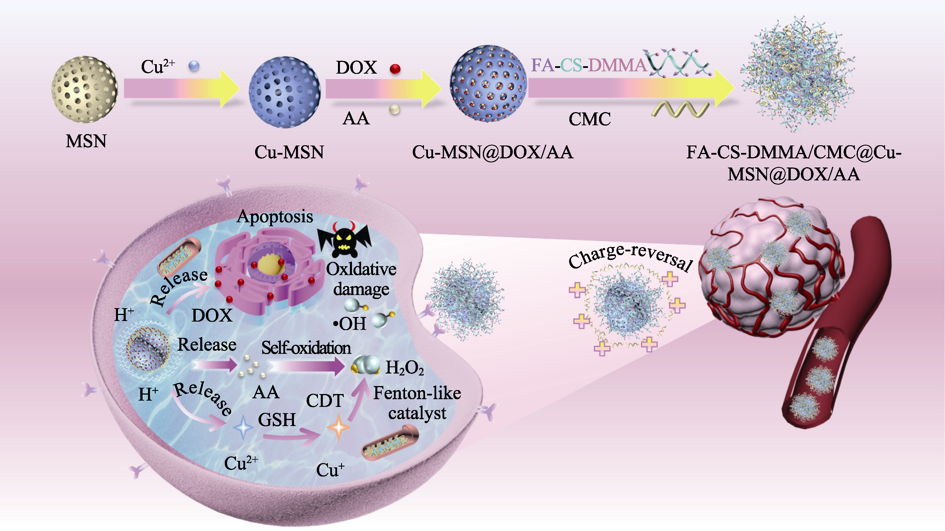
图1 FCDC@Cu-MSN@DA纳米催化剂的制备及增强放大CDT与化疗协同作用的示意图
Fig. 1 Schematic illustration of synthetic procedure and mechanism for enhanced chemo-CDT of nanocatalyst FCDC@Cu-MSN@DA MSN: Mesoporous silica; DOX: Doxorubicin; AA: Ascorbic acid; FA: Folic acid; CS: Chitosan; DMMA: Dimethyl maleic anhydride; CMC: Carboxymethyl chitosan; CDT: Chemodynamic therapy

图2 Cu-MSN和FCDC@Cu-MSN@DA的表征
Fig. 2 Characterization of Cu-MSN and FCDC@Cu-MSN@DA (A, B) SEM images of Cu-MSN (A) and FCDC@Cu-MSN@DA (B); (C, D) Size distributions of Cu-MSN (C) and FCDC@Cu-MSN@DA (D); (E) Elemental (N, O, Si, and Cu) mappings of Cu-MSN; (F) XPS full survey of Cu-MSN; (G) High resolution XPS analysis on Cu2p of Cu-MSN; Colorful figures are available on website
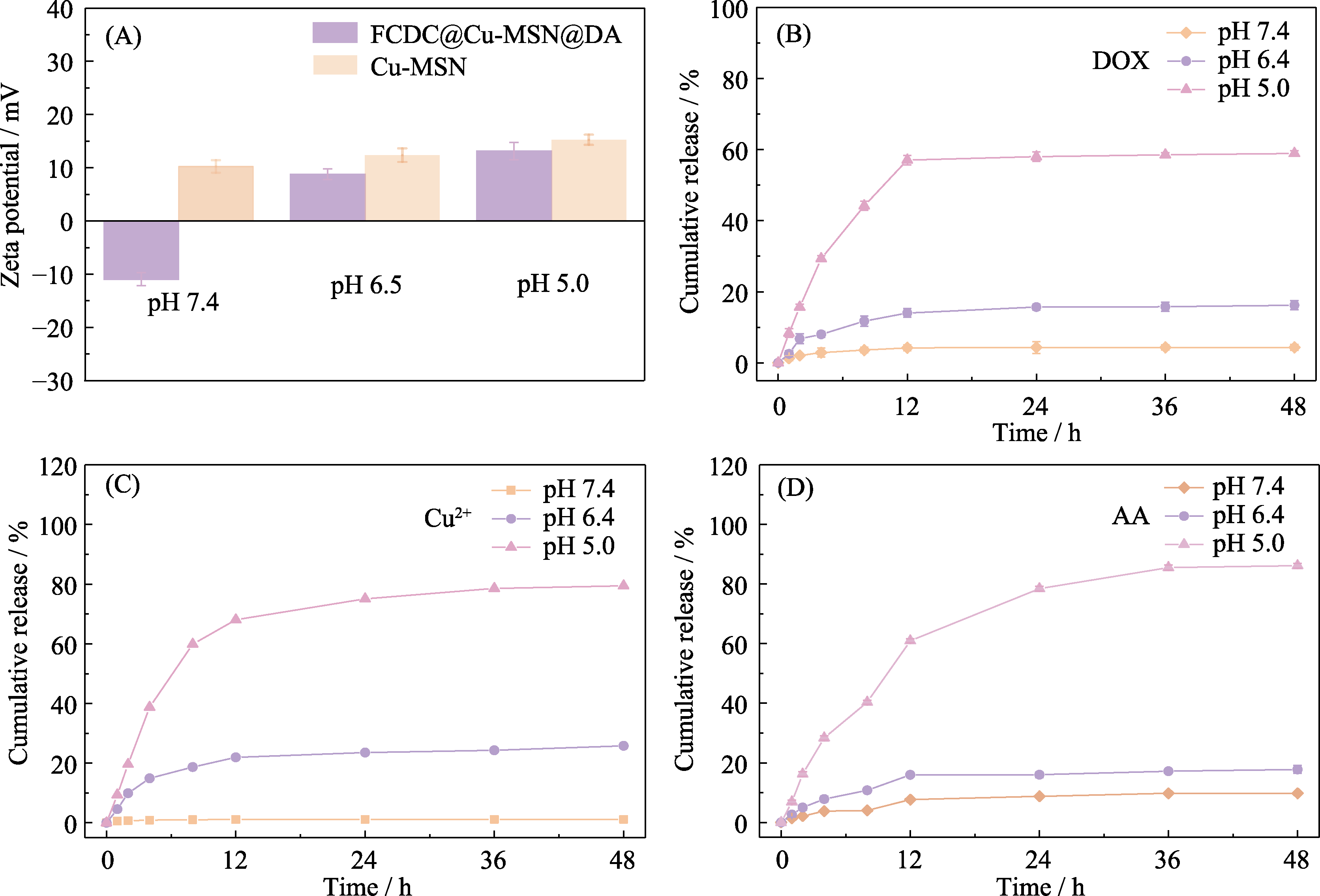
图3 FCDC@Cu-MSN@DA的pH响应特性表征
Fig. 3 pH response of FCDC@Cu-MSN@DA (A) Zeta potential of Cu-MSN and FCDC@Cu-MSN@DA in pH 7.4, 6.5 and 5.0 solutions; (B-D) DOX (B), Cu2+ (C) and AA (D) released from FCDC@Cu-MSN@DA in different pH buffer solutions
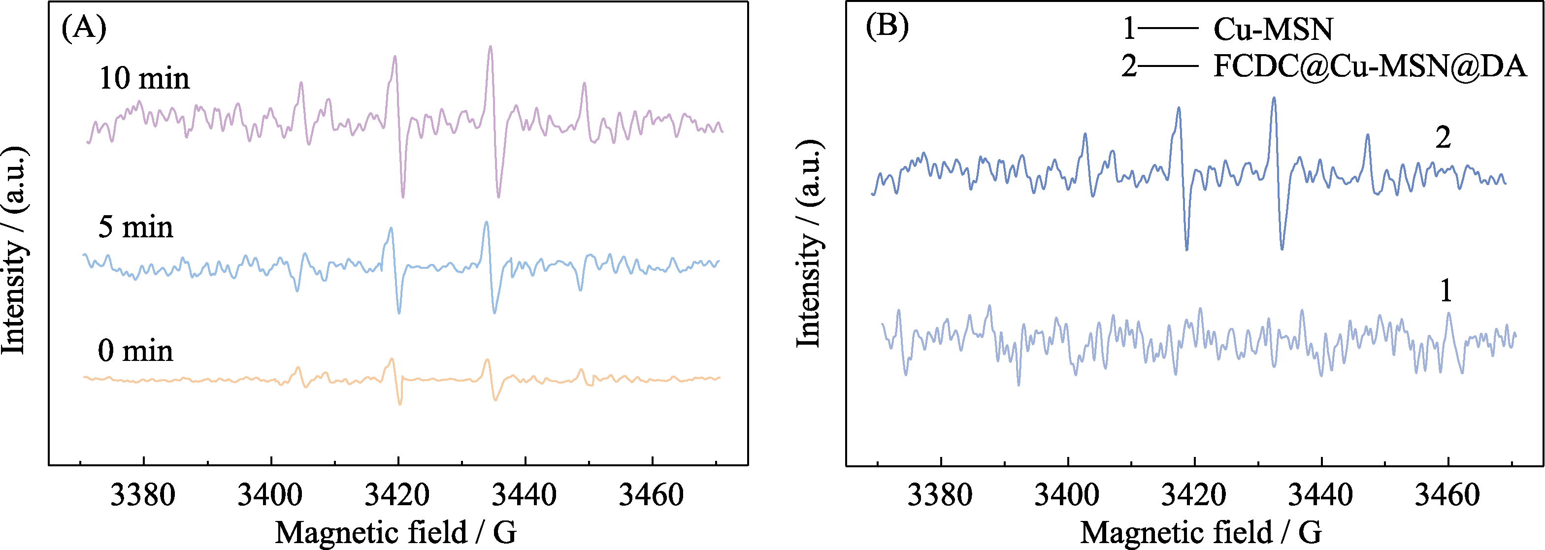
图4 不同催化反应时间的FCDC@Cu-MSN@DA的电子自旋共振能谱(A), Cu-MSN和FCDC@Cu-MSN@DA的电子自旋共振能谱(B)
Fig. 4 ESR signals of FCDC@Cu-MSN@DA for different time (A), ESR spectra of Cu-MSN and FCDC@Cu-MSN@DA (B)
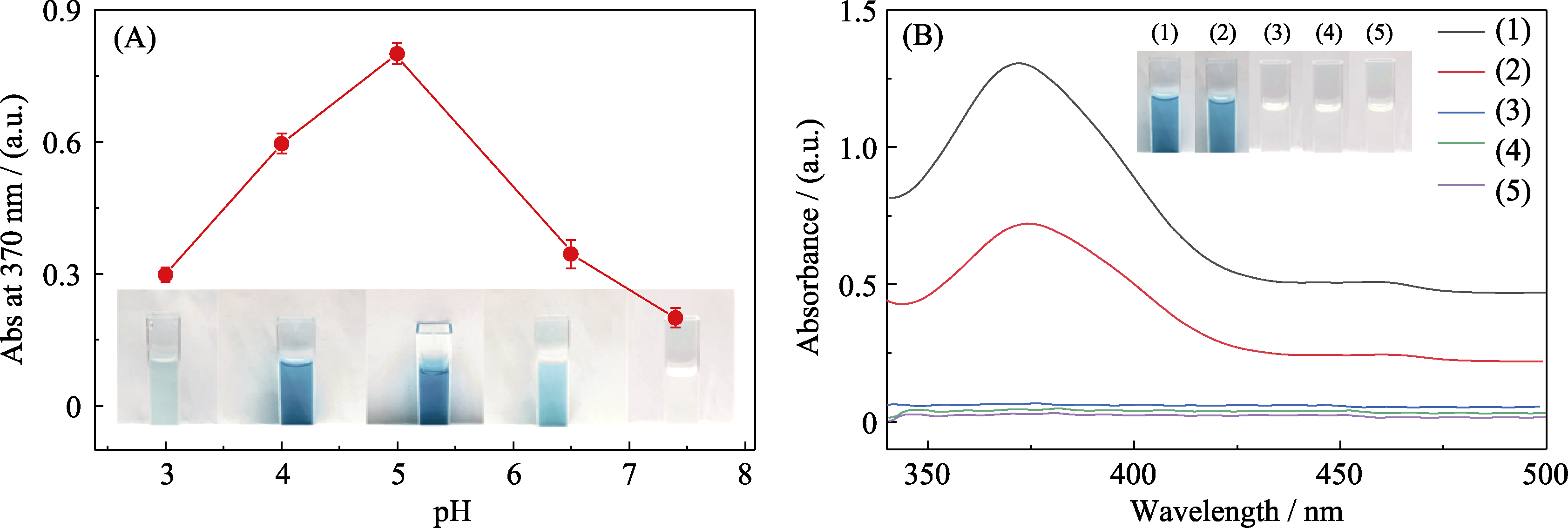
图5 FCDC@Cu-MSN@DA化学动力学性能
Fig. 5 Chemodynamic property of FCDC@Cu-MSN@DA (A) Absorbance of oxidized TMB after treatment with FCDC@Cu-MSN@DA (1 mg/mL) and H2O2 (100 μmol/L) in different pH solutions (pH 3.0, 4.0, 5.0, 6.5, and 7.4); (B) UV-Vis absorption spectra of TMB (oxTMB) catalyzed by (1) TMB+Cu-MSN+H2O2, (2) TMB+FCDC@Cu-MSN@DA, (3) TMB+AA, (4) TMB+H2O2, and (5) TMB in ABS solution (pH 5.0) Insets in (A) and (B) show the corresponding color changes under each pH; TMB: 3,3,5,5-Tetramethylbenzidine; ABS: Acetate buffer solution; Colorful figures are available on website
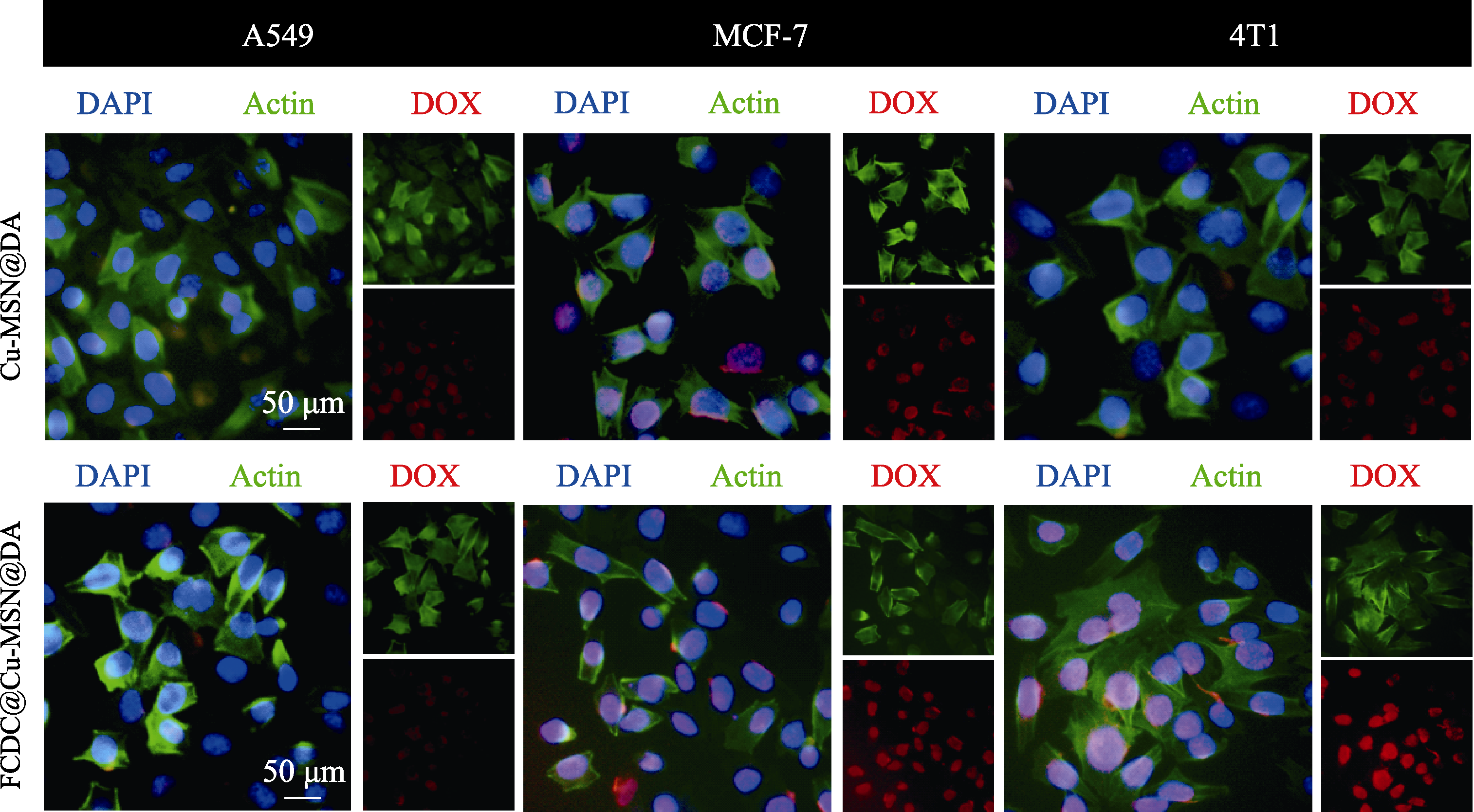
图6 A549、MCF-7 和4T1三种癌细胞吞噬Cu-MSN@DA和FCDC@Cu-MSN@DA的激光共聚焦显微照片
Fig. 6 Laser scanning confocal microscopic images of A549, MCF-7 and 4T1 cancer cells incubated with Cu-MSN@DA and FCDC@Cu-MSN@DA for 5 h DAPI: a staining to show cell neuclei; Actin: a staining to show cell plasm, especially the protein actin; DOX: a staining to show doxolubinson, an anticancer medicine
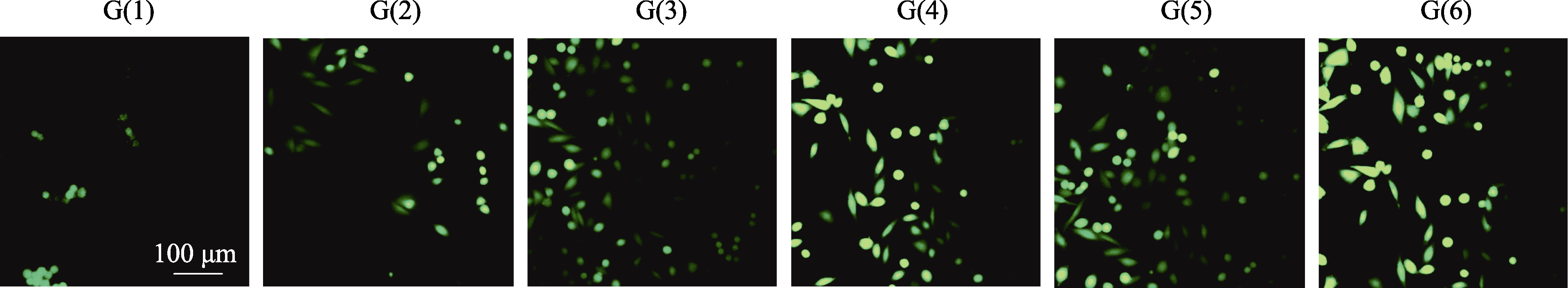
图7 不同样品处理4T1细胞后产生ROS的荧光照片
Fig. 7 Fluorescence images of generation of radical oxigen epecies (ROS) by 4T1 cells incubated with different samples G(1): PBS; G(2): DOX; G(3): FCDC@Cu-MSN@D; G(4): FCDC@Cu-MSN@A; G(5): CDC@Cu-MSN@DA; G(6): FCDC@Cu-MSN@DA
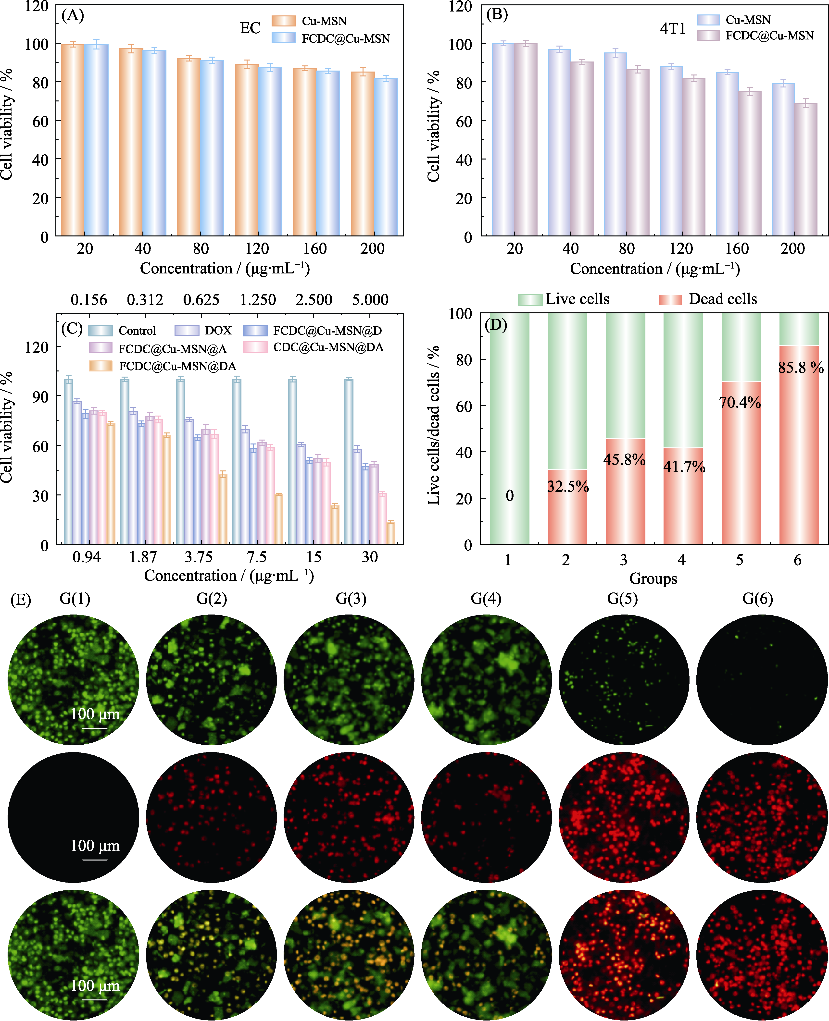
图8 FCDC@Cu-MSN@DA对4T1细胞的化疗-化学动力学协同治疗效果
Fig. 8 Effect of chemo-chemodynamic therapy on 4T1 cells by FCDC@Cu-MSN@DA (A, B) Cell viablity of normal cells (endothelial cell, EC) (A) and cancer cells (4T1 cells) (B) after incubation with different concentrations of Cu-MSN and FCFC@Cu-MSN@DA for 24 h; (C) Cell viability of 4T1 cells incubated with various formulations for 24 h; (D) Semi-quantitative analysis of live/dead cells; (E) Live/dead cell staining images of 4T1 cells after different treatments: G(1): Phosphate buffered saline (PBS); G(2): DOX; G(3): FCDC@Cu-MSN@D; G(4): FCDC@Cu-MSN@A; G(5): CDC@Cu-MSN@DA; G(6): FCDC@Cu-MSN@DA; Colorful figures are available on website
| [1] |
LI Z L, WU H, ZHU J Q, et al. Novel strategy for optimized nanocatalytic tumor therapy: from an updated view. Small Science, 2022, 2(7):2200024.
DOI URL |
| [2] | YANG B W, CHEN Y, SHI J L. Nanocatalytic medicine. Advanced Materials, 2019, 31(39):e1901778. |
| [3] |
WU A, ZHU M, ZHU Y. Copper-incorporated calcium silicate nanorods composite hydrogels for tumor therapy and skin wound healing. Journal of Inorganic Materials, 2022, 37(11):1203.
DOI |
| [4] | ZHU L P, DAI Y L, GAO L Z, et al. Tumor microenvironment- modulated nanozymes for NIR-II-triggered hyperthermia-enhanced photo-nanocatalytic therapy via disrupting ROS homeostasis. International Journal of Nanomedicine, 2021, 16: 4559. |
| [5] |
TANG Z M, ZHAO P R, WANG H, et al. Biomedicine meets Fenton chemistry. Chemical Reviews, 2021, 121: 1981.
DOI URL |
| [6] |
BIAN Y L, LIU B, LIANG S, et al. Cu-based MOFs decorated dendritic mesoporous silica as tumor microenvironment responsive nanoreactor for enhanced tumor multimodal therapy. Chemical Engineering Journal, 2022, 435(2):135046.
DOI URL |
| [7] |
ZHANG W J, ZHAO X Y, LÜ J W, et al. Progresses on hollow periodic mesoporous organosilicas: preparation and application in tumor therapy. Journal of Inorganic Materials, 2022, 37(11):1192.
DOI |
| [8] |
LIU Y, WANG Y H, SONG S Y, et al. Cancer therapeutic strategies based on metal ions. Chemical Science, 2021, 12(37):12234.
DOI PMID |
| [9] | SUN Q Q, WANG Z, LIU B, et al. Recent advances on endogenous/exogenous stimuli-triggered nanoplatforms for enhanced chemodynamic therapy. Coordination Chemistry Reviews, 2022, 451: 214267. |
| [10] | WANG Z, LIU B, SUN Q Q, et al. Fusiform-like copper(II) based metal-organic framework through relief hypoxia and GSH-depletion co-enhanced starvation and chemodynamic synergetic cancer therapy. ACS Applied Materials & Interfaces, 2020, 12(15):17254. |
| [11] |
SHENOY N, CREAGAN E, WITZIG T, et al. Ascorbic acid in cancer treatment: let the phoenix fly. Cancer Cell, 2018, 34(5):700.
DOI PMID |
| [12] | DOSKEY C M, BURANASUDJA V, WAGNER B A, et al. Tumor cells have decreased ability to metabolize H2O2: implications for pharmacological ascorbate in cancer therapy. Redox Biology, 2016, 10: 274. |
| [13] |
YANG B W, SHI J L. Ascorbate tumor chemotherapy by an iron- engineered nanomedicine catalyzed tumor-specific pro-oxidation. Journal of the American Chemical Society, 2020, 142(52):21775.
DOI URL |
| [14] |
AI Y J, SUN H, GAO Z X, et al. Dual enzyme mimics based on metal-ligand cross-linking strategy for accelerating ascorbate oxidation and enhancing tumor therapy. Advanced Functional Materials, 2021, 31(40):2103581.
DOI URL |
| [15] | WU M Q, DING Y M, LI L L. Recent progress in the augmentation of reactive species with nanoplatforms for cancer therapy. Nanoscale, 2019, 11: 19658. |
| [16] | FANG C, DENG Z, CAO G, et al. Co-ferrocene MOF/glucose oxidase as cascade nanozyme for effective tumor therapy. Advanced Functional Materials, 2020, 30: 1910058. |
| [17] |
ZHANG C Y, YAN L, WANG X, et al. Tumor microenvironment-responsive Cu2(OH)PO4 nanocrystals for selective and controllable radiosentization via the X-ray-triggered Fenton-like reaction. Nano Letters, 2019, 19(3):1749.
DOI URL |
| [18] | DONG S M, DONG Y S, JIA T, et al. GSH depleted nanozymes with hyperthermia-enhanced dual enzyme-mimic activities for tumor nanocatalytic therapy. Advanced Materials, 2020, 32(42):e2002439. |
| [19] |
CHEN T, ZENG W W, LIU Y Q, et al. Cu-doped polypyrrole with multi-catalytic activities for sono-enhanced nanocatalytic tumor therapy. Small, 2022, 18(29):2270152.
DOI URL |
| [20] |
NIU J X, SUN S, LIU P F, et al. Copper-based nanozymes: properties and applications in biomedicine. Journal of Inorganic Materials, 2023, 38(5):489.
DOI |
| [21] | XU W J, WANG Y P, HOU G H, et al. Tumor microenvironment responsive hollow nanoplatform for triple amplification of oxidative stress to enhance cuproptosis-based synergistic cancer therapy. Advanced Healthcare Materials, 2023, doi.org/10.1002/adhm.202202949. |
| [22] |
XU W J, QIAN J M, HOU G H, et al. A hollow amorphous bimetal organic framework for synergistic cuproptosis/ferroptosis/apoptosis anticancer therapy via disrupting intracellular redox homeostasis and copper/iron metabolisms. Advanced Functional Materials, 2022, 32(40):2205013.
DOI URL |
| [23] | SHAO L J, HU T S, FAN X Y, et al. Intelligent nanoplatform with multitherapeutic modalities for synergistic cancer therapy. ACS Applied Materials & Interfaces, 2022, 14(11):13122. |
| [24] | MIAO Y L, QIU Y D, YANG W J, et al. Charge reversible and bio-degradable nanocarriers showing dual pH-/reduction-sensitive disinte-gration for rapid site-specific drug delivery. Colloids and Surfaces B: Biointerfaces, 2018, 169: 313. |
| [25] |
TESTA U, PELOSI E, CASTELLI G. New promising developments for potential therapeutic applications of high-dose ascorbate as an anticancer drug. Hematology/Oncology and Stem Cell Therapy, 2021, 14(3):179.
DOI PMID |
| [26] |
LEVINE M, VIOLET P C. Data Triumph at C. Cancer cell, 2017, 31(4):467.
DOI PMID |
| [27] |
WANG Y W, CHEN J J, TIAN Z F, et al. Potassium ferrate-loaded porphyrin-based (VI) metal-organic frameworks for combined photodymanic and chemodynamic tumor therapy. Journal of Inorganic Materials, 2021, 36(12):1305.
DOI |
| [28] |
WU W C, YU L D, JIANG Q Z, et al. Enhanced tumor-specific disulfiram chemotherapy by in situ Cu2+ chelation-initiated nontoxicity-to-toxicity transition. Journal of The American Chemical Society, 2019, 141(29):11531.
DOI URL |
| [29] |
CHENG L C, MA H, SHAO M K, et al. Synthesis of folate-chitosan nanoparticles loaded with ligustrazine to target folate receptor positive cancer cells. Molecular Medicine Reports, 2017, 16(2):1101.
DOI URL |
| [30] |
HONG J Y, SUN Z H, LI Y J, et al. Folate-modified annonaceous acetogenins nanosuspensions and their improved antitumor efficacy. International Journal of Nanomedicine, 2017, 12(1):5053.
DOI PMID |
| [31] | ZHANG L Z, YANG A J, RUAN C P, et al. Copper-nitrogen- coordinated carbon dots: transformable phototheranostics from precise PTT/PDT to post-treatment imaging-guided PDT for residual tumor cells. ACS Applied Materials & Interfaces, 2023, 15 (2): 325. |
| [1] | 汤亚, 孙盛睿, 樊佳, 杨庆峰, 董满江, 寇佳慧, 刘阳桥. 粉煤灰衍生水合硅酸钙PEI改性及吸附去除Cu(II)与催化降解有机污染物[J]. 无机材料学报, 2023, 38(11): 1281-1291. |
| [2] | 陈小梅, 陈颖, 袁霞. 核壳材料Co3O4@SiO2催化环己基过氧化氢分解[J]. 无机材料学报, 2022, 37(1): 65-71. |
| [3] | 马保凯, 李勉, 张绫芷, 翁新楚, 沈彩, 黄庆. 酶-二维MXene复合材料的制备及其电化学检测H2O2的应用[J]. 无机材料学报, 2020, 35(1): 131-138. |
| [4] | 郭露露, 李立霞, 何鹏程, 袁 霞. 介孔材料Co/SBA-15催化环己基过氧化氢分解的研究[J]. 无机材料学报, 2017, 32(5): 543-549. |
| [5] | 郑磊, 李劲, 刘洪波. AEP作用下溶胶-凝胶法制备纤维素基炭气凝胶及其对水溶液中Cu2+吸附性能[J]. 无机材料学报, 2017, 32(11): 1159-1164. |
| [6] | 卢书培, 冯利利, 齐 麟, 王丽丽, 齐兴义. Buserite型氧化锰催化叔丁基过氧化氢歧化分解反应动力学[J]. 无机材料学报, 2016, 31(1): 14-20. |
| [7] | 郑爱芳,陈金龙. CdTe纳米棒的水相合成与铜离子识别研究[J]. 无机材料学报, 2009, 24(2): 251-254. |
| [8] | 章俞之,王忠春,快素兰,胡行方. 锂掺杂MoO3薄膜的制备及电色性能的研究[J]. 无机材料学报, 2000, 15(6): 1131-1135. |
| 阅读次数 | ||||||
|
全文 |
|
|||||
|
摘要 |
|
|||||