Journal of Inorganic Materials ›› 2021, Vol. 36 ›› Issue (10): 1074-1082.DOI: 10.15541/jim20200751
• RESEARCH ARTICLE • Previous Articles Next Articles
YANG Mai1,3( ), ZHU Min1, CHEN Yu2(), ZHU Yufang1,3()
Received:2020-12-31
Revised:2021-02-26
Published:2021-10-20
Online:2021-03-15
Contact:
CHEN Yu, professor. E-mail: chenyuedu@shu.edu.cn; ZHU Yufang, professor. E-mail: zjf2412@163.com
About author:YANG Mai(1996-), female, Master candidate. E-mail: yangmai77@163.com
Supported by:CLC Number:
YANG Mai, ZHU Min, CHEN Yu, ZHU Yufang. FePS3 Nanosheets: Preparation and Potential in Photothermal-photodynamic Therapy[J]. Journal of Inorganic Materials, 2021, 36(10): 1074-1082.
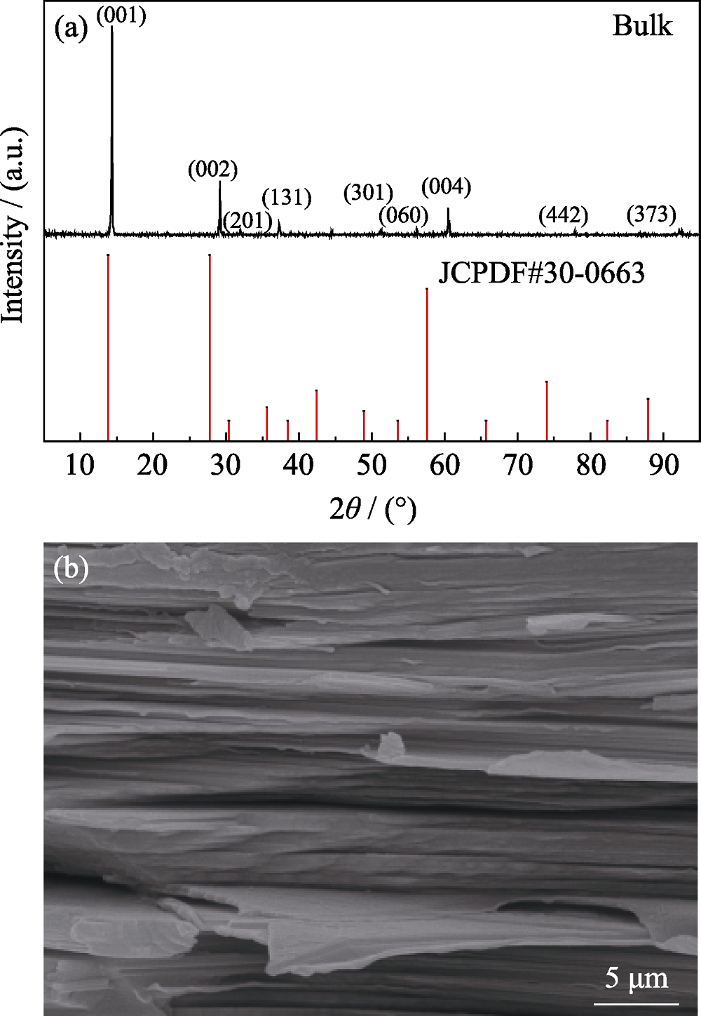
Fig. 1 XRD pattern (a) and SEM image (b) of bulk FePS3
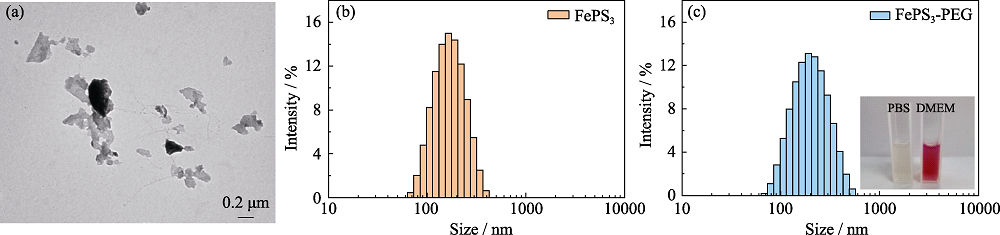
Fig. 2 TEM image (a) of FePS3 nanosheets (NSs) and hydrodynamic size of FePS3 nanosheets (NSs) before (b) and after (c) PEGylation with inset showing the picture of FePS3-PEG dispersed in PBS and in DMEM
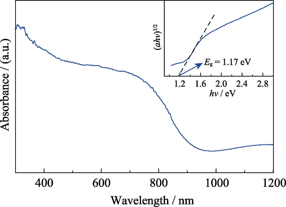
Fig. 3 UV-Vis-NIR diffuse reflectance spectrum of FePS3 NSs with inset showing the estimated band gap potential
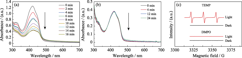
Fig. 4 UV-Vis absorption spectra of the mixture solution of FePS3 NSs mixed with DPBF (1,3-diphenylisobenzofuran) (a) and DPBF solution (b) under 660 nm laser irradiation, and ESR spectra of different reaction systems (c)TEMP: a reagent used to detect 1O2. DMPO: a reagent used to detect O2•- and ∙OH
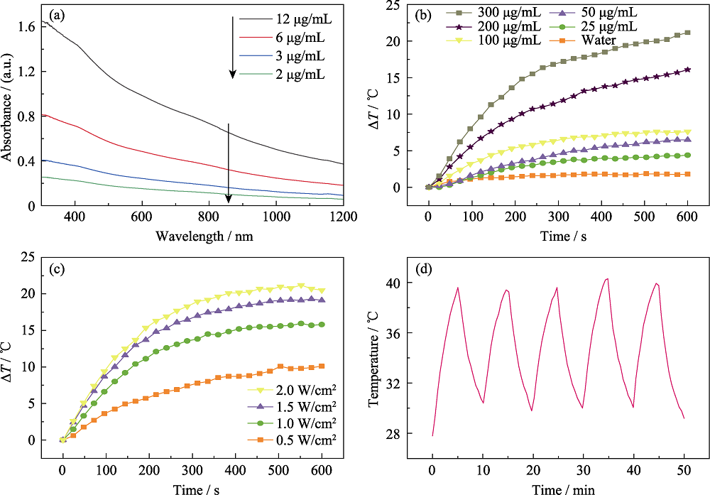
Fig. 5 Vis-NIR spectra of FePS3 NSs with different concentrations (a), photothermal heating curves for different time at different concentrations (b), and different laser power densities (1064 nm laser) (c), and photothermal curve of FePS3 NSs under 5 cycles of laser “on-off” (d)
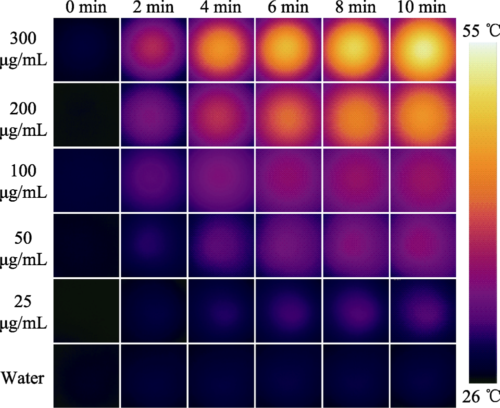
Fig. 6 Thermal images of different concentrations of FePS3 nanosheets heated by 1064 nm laser irradiation for different time
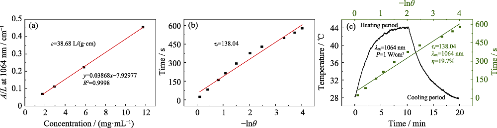
Fig. 7 Linear fitting curve between normalized absorption intensity of FePS3 NSs at λ=1064 nm divided by the characteristic length of the sample at corresponding concentration (A/L) and the corresponding concentration (a), linear relationship between -lnθ and time of cooling process of FePS3 NSs after 1064 nm laser irradiation (b), heating and cooling curves of FePS3 NSs under 1064 nm laser irradiation, and linear relationship between -lnθ and time of the cooling process (c) ε, τs and η represent extinction coefficient, time constant in cooling stage, and photothermal conversion efficiency, respectively, of FePS3 NSs under 1064 nm laser irradiation
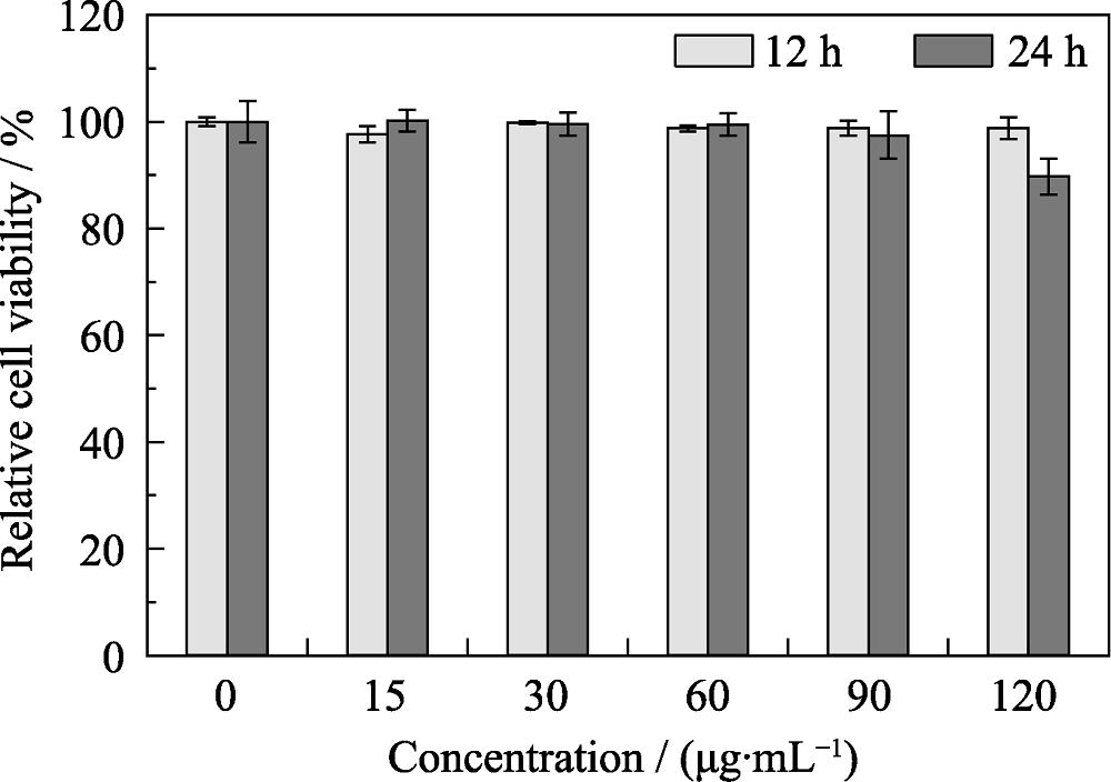
Fig. 8 Relative cell viabilities of 4T1 cells after incubation with different concentrations of FePS3-PEG
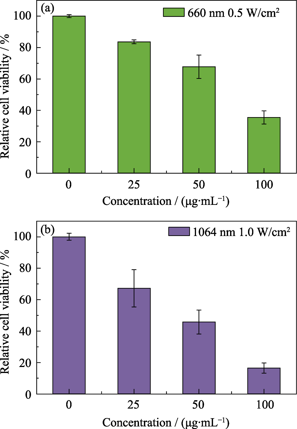
Fig. 9 In vitro photodynamic therapy (a) and photothermal therapy (b) treatment of 4T1 cells after incubation with different concentrations of FePS3-PEG
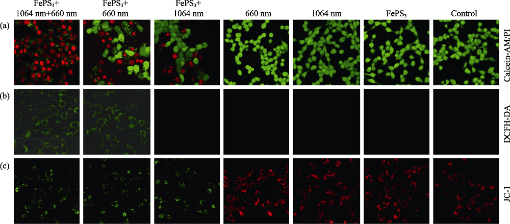
Fig. 10 Confocal laser scanning microscope (CLSM) images of 4T1 cell viabilities (a), reactive oxygen species (ROS) production (b) and changes in mitochondrial membrane potential (c) after different treatments Calcein-AM/PI, DCFH-DA and JC-1 represent methods for detecting cell viability, ROS production, and changes in mitochondrial membrane potential, respectively
| [1] |
LOVELL J F, LIU T W B, CHEN JUAN, et al. Activatable photosensitizers for imaging and therapy. Chemical Reviews, 2010, 110(5):2839-2857.
DOI URL |
| [2] |
CELLI J P, SPRING B Q, RIZVI I, et al. Imaging and photodynamic therapy: mechanisms, monitoring, and optimization. Chemical Reviews, 2010, 110(5):2795-2838.
DOI URL |
| [3] |
DOLMANS D E J G J, FUKUMURA D, JAIN R K. Photodynamic therapy for cancer. Nature Reviews Cancer, 2003, 3(5):380-387.
DOI URL |
| [4] |
PENG B, ANG P K, LOH K P. Two-dimensional dichalcogenides for light-harvesting applications. Nano Today, 2015, 10(2):128-137.
DOI URL |
| [5] |
LI L, KIM J, JIN C, et al. Direct observation of the layer-dependent electronic structure in phosphorene. Nature Nanotechnology, 2016, 12:21-25.
DOI URL |
| [6] |
LI XUAN-HUA, ZHU JIN-MENG, WEI BING-QING. Hybrid nanostructures of metal/two-dimensional nanomaterials for plasmon- enhanced applications. Chemical Society Reviews, 2016, 45:3145-3187.
DOI URL |
| [7] |
NOVOSELOV K S, GEIM A K, MOROZOV S V, et al. Electric field effect in atomically thin carbon films. Science, 2004, 306(5696):666-669.
DOI URL |
| [8] |
CHHOWALLA M, LIU ZHONG-FAN, ZHANG HUA. Two- dimensional transition metal dichalcogenide (TMD) nanosheets. Chemical Society Reviews, 2015, 44(9):2584-2586.
DOI URL |
| [9] |
ALLEN M J, TUNG V C, KANER R B. Honeycomb carbon: a review of graphene. Chemical Reviews, 2009, 110(1):132-145.
DOI URL |
| [10] |
NAGUIB M, KURTOGLU M, PRESSER V, et al. Two-dimensional nanocrystals produced by exfoliation of Ti3AlC2. Advanced Materials, 2011, 23(37):4248-4253.
DOI URL |
| [11] |
LIU HAN, NEAL A T, ZHU ZHEN, et al. Phosphorene: a new 2D material with high carrier mobility. ACS Nano, 2014, 8(4):4033-4041.
DOI URL |
| [12] | MANZELI S, OVCHINNIKOV D, PASQUIER D, et al. 2D transition metal dichalcogenides. Nature Reviews Materials, 2017, 2:17033. |
| [13] | ZHANG XU, ZHAO XU-DONG, WU DI-HUA, et al. MnPSe3 monolayer: a promising 2D visible-light photohydrolytic catalyst with high carrier mobility. Advanced Science, 2016, 3:1600062. |
| [14] | LI XING-XING, WU XIAO-JUN, YANG JIN-LONG. Half-metallicity in MnPSe3 exfoliated nanosheet with carrier doping. Journal of the American Chemical Society, 2014, 136(31):11065-11069. |
| [15] |
MUKHERJEE D, AUSTERIA P M, SAMPATH S. Two-dimensional, few-layer phosphochalcogenide, FePS3: a new catalyst for electrochemical hydrogen evolution over wide pH range. ACS Energy Letters, 2016, 1(2):367-372.
DOI URL |
| [16] |
DU KE-ZHAO, WANG XING-ZHI, LIU YANG, et al. Weak van der Waals stacking, wide-range band gap, and Raman study on ultrathin layers of metal phosphorus trichalcogenides. ACS Nano, 2015, 10(2):1738-1743.
DOI URL |
| [17] | ZHANG QIU-HONG, GUO QIANG-BING, CHEN QIAN, et al. Highly efficient 2D NIR-II photothermal agent with fenton catalytic activity for cancer synergistic photothermal-chemodynamic therapy. Advanced Science, 2020, 7(7):1902576. |
| [18] | FANG XUE-YANG, WU XIAN-LIN, LI ZHEN-DONG, et al. Biomimetic anti-PD-1 peptide-loaded 2D FePSe3 nanosheets for efficient photothermal and enhanced immune therapy with multimodal MR/PA/thermal imaging. Advanced Science, 2020, 8(2):2003041. |
| [19] |
CHENG LIANG, LIU JING-JING, GU XING, et al. PEGylated WS2 nanosheets as a multifunctional theranostic agent for in vivo dual-modal CT/photoacoustic imaging guided photothermal therapy. Advanced Materials, 2014, 26(12):1886-1893.
DOI URL |
| [20] | LIN HAN, GAO SHAN-SHAN, DAI CHEN, et al. Two-dimensional biodegradable niobium carbide (MXene) for photothermal tumor eradication in NIR-I and NIR-II bio-windows. Journal of the American Chemical Society, 2017, 139(45):16235-16247. |
| [21] |
COLEMAN J N, LOTYA M, O’NEILL A, et al. Two-dimensional nanosheets produced by liquid exfoliation of layered materials. Science, 2011, 331(6017):568-571.
DOI URL |
| [22] |
ZHAO WEI, LI AI-HUA, ZHANG AI-TANG, et al. Recent advances in functional-polymer-decorated transition-metal nanomaterials for bioimaging and cancer therapy. ChemMedChem, 2018, 13(20):2134-2149.
DOI URL |
| [23] | ZHANG YONG-CAI, DU ZHEN-NI, LI KUN-WEI, et al. High- performance visible-light-driven SnS/SnO nanocomposite photocatalyst prepared via in situ hydrothermal oxidation of SnS nanoparticles. ACS Applied Materials & Interfaces, 2011, 3(5):1528-1537. |
| [24] | CHENG ZHONG-ZHOU, WANG FENG-MEI, SHIFA T A, et al. Efficient photocatalytic hydrogen evolution via band alignment tailoring: controllable transition from type-I to type-II. Small, 2017, 13(41):1702163. |
| [25] | CHEN PENG, SU YUN, LIU HONG, et al. Interconnected tin disulfide nanosheets grown on graphene for Li-ion storage and photocatalytic applications. ACS Applied Materials & Interfaces, 2013, 5(22):12073-12082. |
| [26] |
DETTY M R, GIBSON S L, WAGNER S J. Current clinical and preclinical photosensitizers for use in photodynamic therapy. Journal of Medicinal Chemistry, 2004, 47(16):3897-3915.
DOI URL |
| [27] |
SHARMAN W M, ALLEN C M, VAN LIER J E . Photodynamic therapeutics: basic principles and clinical applications. Drug Discovery Today, 1999, 4(11):507-517.
DOI URL |
| [28] | WANG HUI, YANG XIAN-ZHU, SHAO WEI, et al. Ultrathin black phosphorus nanosheets for efficient single oxygen generation. Journal of the American Chemical Society, 2015, 137(35):11376-11382. |
| [29] |
LIANG CHEN, ZHANG XING-LIN, YANG MENG-SU, et al. Remodeling tumor microenvironment by multifunctional nanoassemblies for enhanced photodynamic cancer therapy. ACS Materials Letters, 2020, 2(10):1268-1286.
DOI URL |
| [30] | DING XIAN-GUANG, LIOW C H, ZHANG MENG-XIN, et al. Surface plasmon resonance enhanced light absorption and photothermal therapy in the second near-infrared window. Journal of the American Chemical Society, 2014, 136(44):15684-15693. |
| [31] |
BASHKATOV A N, GENINA E A, KOCHUBEY V I, et al. Optical properties of human skin, subcutaneous and mucous tissues in the wavelength range from 400 to 2000 nm. Journal of Physics D: Applied Physics, 2005, 38(15):2543.
DOI URL |
| [32] |
ROBINSON J T, TABAKMAN S M, LIANG YONG-YE, et al. Ultrasmall reduced graphene oxide with high near-infrared absorbance for photothermal therapy. Journal of the American Chemical Society, 2011, 133(17):6825-6831.
DOI URL |
| [33] | LI K C, CHU H C, LIN Y, et al. PEGylated copper nanowires as a novel photothermal therapy agent. ACS Applied Materials & Interfaces, 2016, 8(19):12082-12090. |
| [34] |
SUN CAI-XIA, WEN LING, ZENG JIAN-FENG, et al. One-pot solventless preparation of PEGylated black phosphorus nanoparticles for photoacoustic imaging and photothermal therapy of cancer. Biomaterials, 2016, 91:81-89.
DOI URL |
| [35] |
GIORGIO M, MIGLIACCIO E, ORSINI F, et al. Electron transfer between cytochrome c and p66Shc generates reactive oxygen species that trigger mitochondrial apoptosis. Cell, 2005, 122(2):221-233.
DOI URL |
| [36] |
DANIAL N N, KORSMEYER S J. Cell death: critical control points. Cell, 2004, 116(2):205-219.
DOI URL |
| [37] | WANG YI, WEI GUO-QING, ZHANG XIAO-BIN, et al. Multistage targeting strategy using magnetic composite nanoparticles for synergism of photothermal therapy and chemotherapy. Small, 2018, 14(12):1702994. |
| [1] | BAI Zhiqiang, ZHAO Lu, BAI Yunfeng, FENG Feng. Research Progress on MXenes: Preparation, Property and Application in Tumor Theranostics [J]. Journal of Inorganic Materials, 2022, 37(4): 361-375. |
| Viewed | ||||||
|
Full text |
|
|||||
|
Abstract |
|
|||||