), 郭乐2, 丁嘉仪2, 周嘉琪1, 张学良1, 努尔尼沙·阿力甫1()
), GUO Le2, DING Jiayi2, ZHOU Jiaqi1, ZHANG Xueliang1, NUERNISHA Alifu1()
), 郭乐2, 丁嘉仪2, 周嘉琪1, 张学良1, 努尔尼沙·阿力甫1()
), GUO Le2, DING Jiayi2, ZHOU Jiaqi1, ZHANG Xueliang1, NUERNISHA Alifu1()
图8. UCNPs用于UCL/MRI双模态成像[
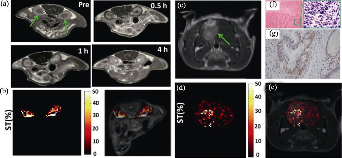
Fig. 8. Application of UCNPs in UCL/MRI dual-modality imaging[
(a) In vivo T1-weighted MRI of kidneys of mice (as arrowed) before and after the intravenous administration of NaGdF4@poly-L-lysine (PLL) nanoparticles; (b) Chemical exchange saturation transfer (CEST) contrast difference map between pre/post injection following radio frequency (RF) irradiation at 3.0 μT, showing the kidney signal in color on the grayscale image to highlight the effect; (c) In vivo T1-weighted MRI of brain tumor (as arrowed) after the intravenous administration of NaGdF4@PLL nanoparticles; (d) CEST contrast difference map between pre/post injection at 3.0 μT, showing the brain ventricle signal in color on the grayscale image to highlight the effect; (e) Merged image of (c, d); (f) H&E staining of the brain tumor tissue; (g) Immunohistochemical staining of the brain tumor tissue, showing the positive expression of glial fibrillary acidic protein