磷酸钙微球骨修复材料研究进展
 , 徐文峰, 廖晓玲
 Research Progress in Calcium Phosphate Microspheres for Bone Defect Repair
LI Bo, XU Wen-Feng, LIAO Xiao-Ling
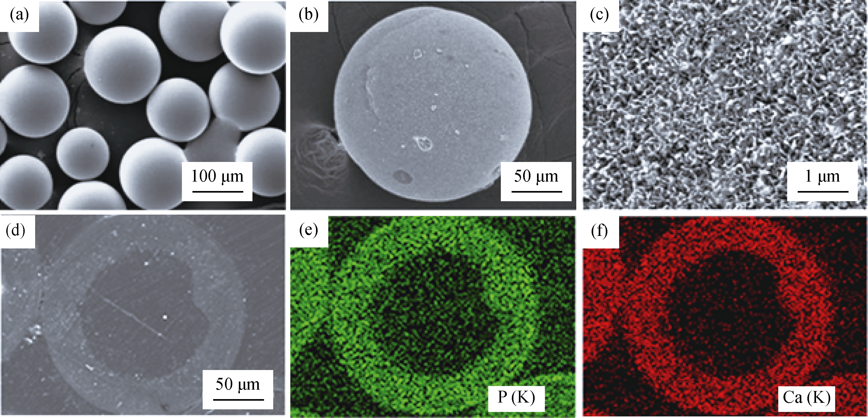
, XU Wen-Feng, LIAO Xiao-Ling Fig. 4 SEM images of hollow HA microsphere with glass as hard template. a starting glass microspheres as hard template, b external surface of hollow HA microsphere, c external surface of hollow HA microsphere at high magnification. d SEM image in back-scattered mode of a polished cross section of a hollow hydroxyapatite microsphere, e and f X-ray maps of CaK and PK across the planar section shown in d [ 27 ]