This is an open-access article distributed under the terms of the Creative Commons Attribution License, which permits unrestricted use, distribution, and reproduction in any medium, provided the original author and source are credited.
Progress on Biomedical Ceramic Coatings Prepared by Thermal Spraying
ZHENG Xue-Bin, XIE You-Tao
Key Laboratory of Inorganic Coating Materials, Shanghai Institute of Ceramics, Chinese Academy of Sciences, Shanghai 200050, China
Fund:National Natural Science Foundation of China (81071455, 51172264);
Abstract
Biomedical ceramic coatings on metallic substrates depositedvia thermal spraying have attracted extensive attentions in the field of orthopedic implants due to their excellent mechanical strengths and biological properties. The studies on biomedical coatings for orthopedic implants are overviewed, and improvements of thermal sprayed hydroxyapatite coatings are summarized. Newly developed bioactive calcium silicate coatings are reviewed as well.
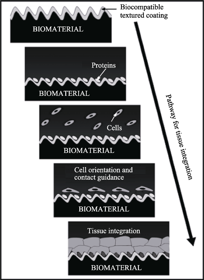
图1 生物材料植入生命系统后发生的系列反应示意图[ 3]Fig. 1 Schematic illustration of the sequential reactions that taken place after the implantation of a biomaterial into a living system[ 3]
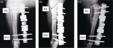
图3 抗菌HA3涂层和常规HA涂层螺钉植入狗胫骨后的X射线照片[ 68]Fig. 3 X-ray views of antibacterial and convenient HA coated screws after implanted in dog’s tibia[ 68](a)1 w, (b)3 w(黑色箭头所指为骨膜炎部位), (c)6 w(白色箭头所指为钉痕)
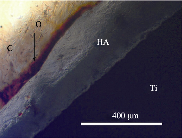
图4 加涂纳米HA涂层的钛植入鼠股骨2 w后的光学显微照片[ 30]Fig. 4 Optical photomicrograph of a longitudinal section of nano-HA coated Ti implanted in rat femur at 2 w showing nonmineralized osteoid (O), and collagen matrix (C)[ 30]
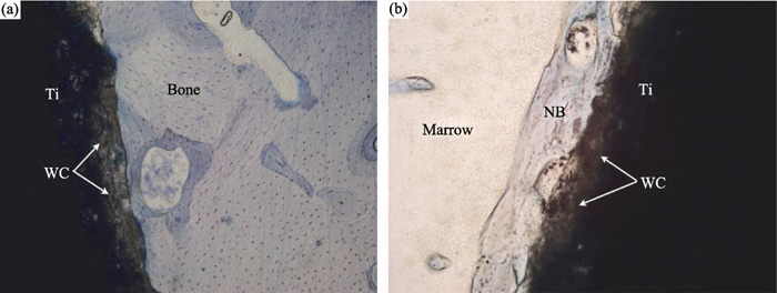
图5 硅灰石涂层在植入股骨(a)与骨髓(b)三个月后的组织学形貌[ 90]Fig. 5 Histological morphologies of the cross-section of the wollastonite coating after implantation in cortical bone (a) and marrow (b) for 3 months[ 90]WC: wollastonite coating, NB: newly formed bone
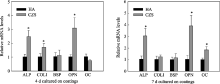
图6 人骨髓基质干细胞(hBMSCs)在CaO-ZrO2-SiO2和HA涂层表面的成骨标志基因表达: (A)4 d, (B)7 d[ 99]Fig. 6 Expression levels of osteoblastic differentiation related genes of hBMSCs cultured on CaO-ZrO2-SiO2 and HA coatings for (A) 4 d and (B) 7 d[ 99]
KasemoB, LausmaaJ. Surface properties and processes of the biomaterial-tissue interface. Materials Science and Engineering C-Biommetic Materials Sensors and and Systems, 1994, 1(3): 115-119. [本文引用:1]
[3]
PaitalS R, DahotreN B. Calcium phosphate coatings for bio-implant applications: Materials, performance factors, and methodologies. Materials Science and Engineering R-Reports, 2009, 66(1/2/3): 1-70. [本文引用:1][JCR: 13.902]
[4]
GittensR A, McLachlanT, Olivares-NavarreteR, et al. The effects of combined micron-/submicron-scale surface roughness and nanoscale features on cell proliferation and differentiation. Biomaterials, 2011, 32(13): 3395-3403. [本文引用:1][JCR: 7.604]
[5]
HatanoK, InoueH, KojoT, et al. Effect of surface roughness on proliferation and alkaline phosphatase expression of rat calvarial cells cultured on polystyrene. Bone, 1999, 25(4): 439-445. [本文引用:1][JCR: 3.823]
[6]
BuserD, SchenkR K, SteinemannS, et al. Influence of surface characteristics on bone integration of titanium implants: a hostomorphometric study in miniature pigs. Journal of Biomedical Materials Research, 1991, 25(7): 889-902. [本文引用:1][JCR: 2.308]
[7]
KellerJ C, YoungF A, HanselB, et al. Systemic effects of porous Ti dental implants. Dental Materials, 1985, 1(2): 41-42. [本文引用:1][JCR: 3.773]
[8]
AebilN, KrebsJ, StichH, et al. In vivo comparison of the osseointegration of vacuum plasma sprayed titanium- and hydroxyapatite-coated implants. Journal of Biomedical Materials Research, 2003, 66A(2): 356-363. [本文引用:2][JCR: 2.308]
[9]
SHIJian-Min, DINGChuan-Xian, WUYi-Hua. Bioactivity of titanium coating. Journal of Inorganic Materials, 2001, 16(3): 515-521. [本文引用:1][JCR: 0.531][CJCR: 1.106]
[10]
ChenY K, ZhengX B, JiH, et al. Effect of Ti-OH formation on bioactivity of vacuum plasma sprayed titanium coating after chemical treatment. Surface & Coating Technology, 2007, 202(3): 494-498. [本文引用:1]
[11]
XueW C, LiuX Y, ZhengX B, et al. In vivo evaluation of plasma sprayed titanium coating after alkali modification. Biomaterials, 2005, 26(16): 3029-3037. [本文引用:1][JCR: 7.604]
[12]
SchrootenJ, HesenJ A. Adhesion of bioactive glass coating to Ti6Al4V oral implant. Biomaterials, 2000, 21(14): 1461-1469. [本文引用:1][JCR: 7.604]
[13]
WangG C, LiuX Y, GaoJ H, et al. In vitro bioactivity and phase stability of plasma-sprayed nanostructured 3Y-TZP coatings. Acta Biomaterialia, 2009, 5(6): 2270-2278. [本文引用:1][JCR: 5.093]
[14]
HanY, ChenD, SunJ, et al. UV-enhanced bioactivity and cell response of micro-arc oxidized titania coatings. Acta Biomaterialia, 2008, 4(5): 1518-1529. [本文引用:1][JCR: 5.093]
[15]
FeddesB, VredenbergA M, WolkeJ G C, et al. Bulk composition of r. f. magnetron sputter deposited calcium phosphate coatings on different substrates (polyethylene, polytetrafluoroethylene, silicon). Surface & Coatings Technology, 2004, 185(2/3): 346-355. [本文引用:1]
[16]
GuehennecL, Lopez-HerediaM A, EnkelB, et al. Osteoblastic cell behaviour on different titanium implant surfaces. Acta Biomaterialia, 2008, 4(3): 535-543. [本文引用:1][JCR: 5.093]
FroimsonM I, GarinoJ, MachenaudA, et al. Minimum 10-year results of a tapered, titanium, hydroxyapatite-coated hip stem: an independent review. The Journal of Arthroplasty, 2007, 22(1): 1-7. [本文引用:1]
[19]
YuL G, KhorK A, LiH, et al. Effect of spark plasma sintering on the microstructure and in vitro behavior of plasma sprayed HA coatings. Biomaterials, 2003, 24(16): 2695-2705. [本文引用:1][JCR: 7.604]
[20]
DaugaardH, ElmengaardB, BechtoldJ E, et al. The effect on bone growth enhancement of implant coatings with hydroxyapatite and collagen deposited electrochemically and by plasma spray. Journal of Biomedical Materials Research, 2010, 92A(3): 913-921. [本文引用:1][JCR: 2.308]
[21]
GrootK, GeesinkR G T, KleinC P A T, et al. Plasma sprayed coating of hydroxyapatite. Journal of Biomedical Materials Research, 1987, 21(12): 1375-1387. [本文引用:1][JCR: 2.308]
[22]
LacefieldW R. Hydroxyapatite coatings, In: An Introduction to Bioceramics, ed. Hench L L and Wilson J. Singapore: World Scientific, 1994. [本文引用:1]
CheangP, KhorK A. Addressing processing problems associated with plasma spraying hydroxyapatite coatings. Biomaterials, 1996, 17(5): 537-544. [本文引用:2][JCR: 7.604]
[25]
FengC F, KhorK A, LiuE J, et al. Phase transformations in plasma sprayed hydroxyapatitecoatings. Scripta Materialia, 1999, 42(1): 103-109. [本文引用:1][JCR: 2.821]
[26]
ZHENGXue-Bin, HUANGMin-Hui, HUANGJing-Qi, et al. Effect of spray distance and spray power on properties of plasma sprayed hydroxyapatite coatings. Journal of Inorganic Materials, 1999, 14(5): 783-788. [本文引用:3][JCR: 0.531][CJCR: 1.106]
[27]
DongZ L, KhorK A, QuekC H, et al. TEM and STEM analysis on heat-treated and in vitro plasma-sprayed hydroxyapatite/ Ti-6Al-4V composite coatings. Biomaterials, 2003, 24(1): 97-105. [本文引用:1][JCR: 7.604]
[28]
CizekJ, KhorK A. Role of in-flight temperature and velocity of powder particles on plasma sprayed hydroxyapatite coating characteristics. Surface & Coatings Technology, 2012, 206(8/9): 2181-2191. [本文引用:1]
[29]
BhadangK A, GrossK A. Influence of fluorapatite on the properties of thermally sprayed hydroxyapatite coatings. Biomaterials, 2004, 25(20): 4935-4945. [本文引用:1][JCR: 7.604]
[30]
RoyM, Band yopadhyayA, BoseS, et al. Induction plasma sprayed nano hydroxyapatite coatings on titanium for orthopaedic and dental implants. Surface & Coatings Technology, 2011, 205(8/9): 2785-2792. [本文引用:1]
[31]
BrossaF, CigadaA, ChiesaR, et al. Post-deposition treatment effects on hydroxyapatite vacuum plasma spray coatings. Journal of Materials Science - Materials in Medicine, 1994, 5(12): 855-857. [本文引用:2][JCR: 2.141]
[32]
YangY C. Influence of residual stress on bonding strength of the plasma-sprayed hydroxyapatite coating after the vacuum heat treatment. Surface & Coatings Technology, 2007, 201(16/17): 7187-7193. [本文引用:1]
[33]
TaoS Y, JiH, DingC X. Effect of vapor-flame treatment on plasma sprayed hydroxyapatite coatings. Journal of Biomedical Materials Research, 2000, 52(3): 572-575. [本文引用:1][JCR: 2.308]
[34]
XueW C, LiuX Y, ZhengX B, et al. Effect of hydroxyapatite coating crystallinity on dissolution and osseointegration in vivo. Journal of Biomedical Materials Research Part A, 2005, 74A(4): 553-561. [本文引用:1]
[35]
GrossK A, Rodriguez-LorenzoL M. Sintered hydroxyfluorapatites. Part I: Sintering ability of precipitated solid solution powders. Biomaterials, 2004, 25: 1375-1384. [本文引用:1][JCR: 7.604]
[36]
RobinsonC, ShoreR C, BrookesS J, et al. The chemistry of enamel caries. Crit. Rev. Oral. Biol. Med. , 2000, 11(4): 481-495. [本文引用:1]
[37]
Land iE, TampieriA, Mattioli-Belmonte, et al. Biomimetic Mg- and Mg, CO32- substituted hydroxyapatites: synthesis characterization and in vitro behavior. J. Eur. Ceram. Soc. , 2006, 26(13): 2593-2601. [本文引用:1][JCR: 2.36]
Land iE, TampieriA, CelottiG, et al. Sr-substituted hydroxyapatites for osteoporotic bone replacement. Acta Biomater. , 2007, 3(6): 961-969. [本文引用:1][JCR: 5.093]
[40]
OliveiraA L, ReisR L, LiP. Strontium-substituted apatite coating grown on Ti6Al4V substrate through biomimetic synthesis. J. Biomed. Mater. Res. B, 2007, 83(1): 258-265. [本文引用:1][JCR: 2.147]
[41]
XueW, HosickH L, Band yopadhyayA, et al. Preparation and cell-materials interactions of plasma sprayed strontium-containing hydroxyapatite coating. Surf. Coat. Technol. , 2007, 201(8): 4685-4693. [本文引用:1][JCR: 1.941]
[42]
SchwarzK, MilneD. Growth-promoting effects of silicon in rats. Nature, 1972, 239: 333-334. [本文引用:1][JCR: 38.597]
[43]
CarlisleE. Silicon: a possible factor in bone calcification. Science, 1970, 167(3916): 279-280. [本文引用:1]
[44]
PietakA M, ReidJ W, StottM J, et al. Silicon substitution in the calcium phosphate bioceramics. Biomaterials, 2007, 28: 4023-4032. [本文引用:1][JCR: 7.604]
[45]
JugdaohsinghR, TuckerK, QiaoN, et al. Dietary silicon intake is positevely associated with bone mineral density in men and premenopausal women of the Framingham offspring cohort. J. Bone. Miner. Res. , 2004, 19(2): 297-307. [本文引用:1][JCR: 6.128]
[46]
DarleyW M, VolcaniB E. Role of silicon in diatom metabolism. A silicon requirement for deoxyreibonucleic acid synthesis in diatom Cylindrotheca-fusiformis reimann and lewin. Exp. Cell. Res. , 1969, 58(2/3): 334. [本文引用:1][JCR: 3.557]
[47]
KeetingP, OurslerM, Wiegand K, et al. Zeolite a increases proliferation, differentiation, and transforming growth factor beta production in normal adult human osteoblastlike cells in vitro. J. Bone. Miner. Res. , 1992, 7(11): 1281-1289. [本文引用:1][JCR: 6.128]
[48]
ReffittD, OgstonN, JugdaohsinghR, et al. Orthosilicic acid stimulates collagen type I synthesis and osteoblastic differentiation in human osteoblastic-like cells in vitro. Bone, 2003, 32(2): 127-135. [本文引用:1][JCR: 3.823]
GuY W, KhorK A, CheangP. In vitro studies of plasma-sprayed hydroxyapatite/Ti-6Al-4V composite coatings in simulated body fluid (SBF). Biomaterials, 2003, 24(9): 1603-1611. [本文引用:1][JCR: 7.604]
[51]
KhorK A, GuY W, QuekC H, et al. Plasma spraying of functionally graded hydroxyapatitey/Ti-6Al-4V coatings. Surface & Coatings Technology, 2003, 168(2/3): 195-201. [本文引用:2]
[52]
LimV J P, KhorK A, FuL, et al. Hydroxyapatite-zirconia composite coatings via the plasma spraying process. Journal of Materials Processing Technology, 1999, 89-90(SI): 491-496. [本文引用:1][JCR: 1.953]
[53]
LiH, KhorK A, KumarR, et al. Characterization of hydroxyapatite nano-zirconia composite coatings deposited by high velocity oxy-fuel (HVOF) spray process. Surface & Coatings Technology, 2004, 182(2/3): 227-236. [本文引用:1]
[54]
ZhengX B, DingC X. Characterization of plasma sprayed hydroxyapatite / zirconium oxide composite coatings. Journal of the Korean Vacuum Society, 2000, 9(SI): 89-94. [本文引用:1]
GristinaA G. Biomaterial-centered infection: microbial adhesion versus tissue integration. Science, 1987, 237(4822): 1588-1595. [本文引用:1]
[59]
GruessnerU, ClemensM, PahlplatzP V, et al. Improvement of peripheral wound healing by local administration of gentamicin- impregnated collagen fleeces after abdominoperineal excision of rectal cancer. American Journal of Surgery, 2001, 182: 502-509. [本文引用:1][JCR: 2.516]
[60]
OyaneA, YokoyamaY, UchidaM, et al. The formation of an antibacterial agent-apatite composite coating on a polymer surface using a metastable calcium phosphate solution. Biomaterials, 2006, 27(17): 3295-3303. [本文引用:1][JCR: 7.604]
[61]
SorensenT S, SorensenA L. Rapid release of gentamycin from collagen sponge: in vitro comparison with plastic beads. Acta Orthopaedice Scand inavica, 1990, 61(4): 353-356. [本文引用:1]
[62]
FrédéricL, AurélienB, JérémyG, et al. A new concept of gentamicin loaded HAP/TCP bone substitute for prophylactic action: in vitro release validation. Journal of Materials Science - Materials in Medicine, 2008, 19(2): 947-951. [本文引用:1][JCR: 2.141]
[63]
CambellA A, SongL, LiX S, et al. Development, characterization, and anti-microbial efficacy of hydroxyapatite-chlorhexidine coatings produced by surface-induced mineralization. Journal of Biomedical Materials Research, 2000, 53(4): 400-407. [本文引用:1][JCR: 2.308]
[64]
FengQ L, KimT N, WuJ, et al. Antibacterial effects of Ag-HAp thin films on alumina substrates. Thin Sold Films, 1998, 335(1/2): 214-219. [本文引用:1]
[65]
WanY Z, XiongG Y, LiangH, et al. Modification of medical metals by ion implantation of copper. Applied Surface Science, 2007, 253(24): 9426-9429. [本文引用:1][JCR: 2.112]
[66]
ChenYikai, ZhengXuebin, XieYoutao, et al. Anti-bacterial and cytotoxic properties of plasma sprayed silver-containing HA coatings. Journal of Materials Science - Materials in Medicine, 2008, 19(12): 3603-3609. [本文引用:1][JCR: 2.141]
TerceroJ E, NaminS, LahiriD, et al. Effect of carbon nanotube and aluminum oxide addition on plasma-sprayed hydroxyapatite coating's mechanical properties and biocompatibility. Materials Science and Engineering C, 2009, 29(7): 2195-2202. [本文引用:1]
[70]
BalaniK, AndersonR, LahaT, et al. Plasma-sprayed carbon nanotube reinforced hydroxyapatite coatings and their interaction with human osteoblasts in vitro. Biomaterials, 2007, 28(4): 618-624. [本文引用:1][JCR: 7.604]
[71]
LiH, KhorK A, CheangP. Adhesive and bending failure of thermal sprayed hydroxyapatite coatings: Effect of nanostructures at interface and crack propagation phenomenon during bending. Engineering Fracture Mechanics, 2007, 74(12): 1894-1903. [本文引用:1][JCR: 1.413]
GadowR, KillingerA, StieglerN. Hydroxyapatite coatings for biomedical applications deposited by different thermal spray techniques. Surface & Coatings Technology, 2010, 205(4): 1157-1164. [本文引用:1]
[74]
HuangY, SongL, HuangT. Characterization and formation mechanism of nano-structured hydroxyapatite coatings deposited by the liquid precursor plasma spraying process. Biomedical Materials, 2010, 5(5): 054113-1-10. [本文引用:1][JCR: 2.158]
[75]
HuangY, SongL, LiuX Y, et al. Hydroxyapatite coatings deposited by liquid precursor plasma spraying: controlled dense and porous microstructures and osteoblastic cells responses. Biofabrication, 2010, 2(4): 045003-1-3. [本文引用:1][JCR: 3.705]
[76]
LiuX Y, ZhaoX, DingC X, et al. Light-induced bioactive TiO2 surface. Applied Physics Letters, 2006, 88(1): 013905. [本文引用:1][JCR: 3.794]
[77]
ZhaoX B, LiuX Y, ChenZ G, et al. Study on bioactivity of plasma-sprayed titania coating. Journal of Inorganic Materials, 2008, 23(5): 1021-1026. [本文引用:1][JCR: 0.531][CJCR: 1.106]
[78]
HuX X, ShenH, ChengY, et al. One-step modification of nano-hydroxyapatite coating on titanium surface by hydrothermal method. Surf Coat Technol, 2010, 205(7): 2000-2006. [本文引用:1][JCR: 1.941]
[79]
WangG, LiuX, ZreiqatH, et al. Enhanced effects of nano-scale topography on the bioactivity and osteoblast behaviors of micron rough ZrO2 coatings. Colloids and Surfaces B: Biointerfaces, 2011, 86(2): 267-274. [本文引用:1][JCR: 3.554]
[80]
LiuX Y, TaoS Y, DingC X. Bioactivity of plasma sprayed dicalcium silicate coating. Biomaterials, 2002, 23(3): 963-968. [本文引用:2][JCR: 7.604]
[81]
LiuX Y, DingC X. Reactivity of plasma sprayed wollastonite in simulated body fluid. J. Biomed. Mater. Res. , 2002, 59(2): 259-264. [本文引用:1][JCR: 2.308]
[82]
LiuX Y, DingC X. Apatite formed on the surface of plasma sprayed wollastonite coating immersed in simulated body fluid. Biomaterials, 2001, 22(14): 2007-2012. [本文引用:1][JCR: 7.604]
[83]
XueW C, LiuX Y, ZhengX B, et al. Dissolution and mineralization of plasma-sprayed wollastonite coatings with different crystallinity. Surf. Coat. Technol, 2004, 200(7): 2420-2427. [本文引用:2][JCR: 1.941]
[84]
XueW C, DingC X, CaoC, et al. Bioactivity of plasma-sprayed diopside coating in vitro. Key Eng. Mater. ,2005, 288-289: 319-322. [本文引用:1][JCR: 0.224]
[85]
LiuX Y, DingC X. Phase compositions and microstructure of plasma sprayed wollastonite coating. Surf. Coat. Technol. , 2001, 141(2/3): 269-274. [本文引用:1][JCR: 1.941]
[86]
LiuX Y, DingC X. Characterization of plasma sprayed wollastonite powder and coating. Surf. Coat. Technol. , 2002, 153(2/3): 173-177. [本文引用:1][JCR: 1.941]
XueW C, LiuX Y, ZhengX B, et al. Plasma-sprayed diopside coatings for biomedical applications. Surf. Coat. Technol. , 2004, 185(2/3): 340-345. [本文引用:1][JCR: 1.941]
[89]
LiuX Y, DingC X. Thermal properties and microstructure of a plasma sprayed wollastonite coating. J. Therm. Spray. Technol. , 2002, 11(3): 375-379. [本文引用:1]
[90]
XueW C, LiuX Y, ZhengX B, et al. In vivo evaluation of plasma-sprayed wollastonite coating. Biomaterials, 2005, 26(17): 3455-3460. [本文引用:1][JCR: 7.604]
[91]
XieY T, LiuX Y, ZhengX B, et al. Bioconductivity of plasma sprayed dicalcium silicate/titanium composite coatings on Ti-6Al-4V alloy. Surf. Coat. Technol. , 2005, 199(1): 105-111. [本文引用:1][JCR: 1.941]
XieY T, LiuX Y, DingC X, et al. Bioconductivity and mechanical properties of plasma-sprayed dicalcium silicate/zirconia composite coatings. Mater. Sci. Eng. , C, 2005, 25(4): 509-515. [本文引用:1][JCR: 2.404]
[94]
XieY T, ChuP K, LiuX Y, et al. Improved stability of dicalcium silicate/zirconia composite coatings by post-spraying heat treatment. Solid State Phenomena, 2005, 107: 141-144. [本文引用:1][JCR: 0.493]
[95]
LiangY, XieY T, JiH, et al. Chemical Stability and biological properties of plasma-sprayed CaO-SiO2-ZrO2 Coatings. J. Therm. Spray Technol. , 2010, 19(6): 1171-1178. [本文引用:1]
[96]
XieY T, ZhengX B, DingC X, et al. Preparation and characterization of CaO-ZrO2-SiO2 coating for potential application in biomedicine. J. Therm. Spray Technol. , 2009, 18(4): 678-685. [本文引用:1]
[97]
LiangY, XieY T, JiH, et al. Excellent stability of plasma-sprayed bioactive Ca3ZrSi2O9 ceramic coating on Ti-6Al-4V. Appl. Surf. Sci. , 2010, 256(14): 4677-4681. [本文引用:1][JCR: 2.112]
YangF, XieY T, LiH, et al. Human bone marrow-derived stromal cells cultured with a plasma sprayed CaO-ZrO2-SiO2 coating. J. Biomed. Mater. Res. B, 2010, 95B(1): 192-201. [本文引用:1][JCR: 2.147]
100
LiK, YuJ, XieY T, et al. Chemical stability and antimicrobial activity of plasma sprayed bioactive Ca2ZnSi2O7 coating. J. Mater. Sci. - Mater. Med. , 2011, 22(12): 2781-2789. [本文引用:1]
1. Shanghai Institute of Ceramics; Chinese Academy of Sciences; Shanghai 200050 China; 2. College of Chemistry and Chemical Technology; Shanghai Jiaotong University; Shanghai; 200240 China
Vacuum plasma sprayed Ti coatings were chemically treated with 5.0mol/L NaOH at 40℃ for 24h and immersed into two kinds of simulated physiological solutions containing Ca2+, HPO2-4 ions. The surface morphologies and compositions of Ti coating before and after alkali treatment were examined by SEM and AES respectively. The formation layers on the Ti coating after immersion in FCS and SBF were characterized by SEM, EDS, XRD and FT-IR. The changes of ion concentration and pH value of FCS after immersion were measured by ICP-AES and pH meter. The results obtained indicate that the fibrous and net-like surface structure forms on the surface of porous Ti coating by treatment of NaOH solution which consists of Na,Ti and O element with the atom ratio nearly equal to that of Na2TiO3. After immersion in simulated physiological solutions, the treated Ti coating shows apatite-forming ability and the apatite formed in FCS is oriented crystallized, plate-like HA with trace of OCP whereas the apatite precipitated in SBF is needle-like carbonate-containing hydroxyapatite. The bioactivity of the alkali treated Ti coatings can be explained in terms of the formation of hydrated titanate which can induced apatite nucleation and growth in FCS and SBF.
In this paper, plasma-sprayed titanium coatings were modified by alkali treatment. The changes in chemical composition and structure of coatings were examined by SEM and AES. The results obtained indicated that a net-like microscopic texture feature, which was full of the interconnected fine porosity, appeared on the surface of alkali-modified titanium coatings. The surface chemical composition was also altered by alkali modification. A sodium titanate compound was formed on the surface of the titanium coating and replaced the native passivating oxide layer. Its thickness was measured as about 150 nm which was about 10 times of that of the as-sprayed coating. The bone bonding ability of titanium coatings were investigated using a canine model. The histological examination and SEM observation demonstrated that more new bone was formed on the surface of alkali-modified implants and grew more rapidly into the porosity. The alkali-modified implants were found to appose directly to the surrounding bone. In contrast, a gap was observed at the interface between the as-sprayed implants and bone. The push-out test showed that alkali-modified implants had a higher shear strength than as-sprayed implants after 1 month of implantation. An interfacial layer, containing Ti, Ca and P, was found to form at the interface between bone and the alkali-modified implant by EDS analysis.
In this work, plasma-sprayed nanostructured zirconia coatings stabilized with 3 mol.% yttria (3Y-TZP) were deposited on Ti substrates. The microstructure and phase composition of coatings were characterized using scanning electron microscopy and X-ray diffraction. The in vitro bioactivity of coatings was evaluated by examining the formation of bone-like apatite on its surface in simulated body fluid. MG63 cell lines were cultured on the coating to investigate its cytocompatibility. The crystalline phase of the as-sprayed coating was tetragonal zirconia, and no monoclinic zirconia was detected. The size of the grains on the as-sprayed coating surface was less than 100 nm. The apatite could precipitate on the surface of the coating immersed in simulated body fluid for 28 days while no apatite was formed on the surface of 3Y-TZP ceramic control, indicating that the bioactivity of the coating is superior to the ceramic with the same composition. It also revealed that the polished coating whose nanostructural outmost layer was removed was bioinert, implying the significance of the nanosized grains for its bioactivity. MG63 cells could adhere, grow and proliferate well on the coating surface, indicating that the coating had good cytocompatibility. Phase stability of plasma-sprayed 3Y-TZP coating was evaluated under hydrothermal conditions at 134 °C. It revealed that the plasma-sprayed nanostructured zirconia coating was more sensitive to aging than that of zirconia ceramics.
Using ultraviolet (UV) irradiation of micro-arc oxidized (MAO) titania coating in distilled water for 0.5 and 2 h, we have achieved an enhanced bioactivity and cell response to titania surface. The MAO coating appears porous and predominantly consists of nanocrystallized anatase TiO2. Compared with the MAO coating, the UV-irradiated coatings do not exhibit any obvious change in surface roughness, morphology, grain size and phase component; however, they have more abundant basic Ti–OH groups and become more hydrophilic because the water contact angle decreases significantly from 17.9 ± 0.8° to 0°. In simulated body fluid (SBF), bonelike apatite-forming ability is significantly stronger on the UV-irradiated coatings than the MAO coating. SaOS-2 human osteoblast-like cell attachment, proliferation and alkaline phosphatase of the cell are greater on the UV-irradiated coatings relative to the MAO coating. UV irradiation of titania results in the conversion of Ti4+ to Ti3+ and the generation of oxygen vacancies, which could react with the absorbed water to form basic Ti–OH groups. The enhanced bioactivity and cell response of the UV-irradiated coatings are proven to result from abundant Ti–OH groups on the coating surfaces. After storing the UV-irradiated coatings in the dark for two weeks, the basic Ti–OH groups on the coatings slightly decrease in amount and can induce apatite formation after a short period of SBF immersion, and show relative long-term stability.
The osseointegration of oral implants is related to the early interactions between osteoblastic cells and titanium surfaces. The behaviour of osteoblastic MC3T3-E1 cells was compared on four different titanium surfaces: mirror-polished (Smooth-Ti), alumina grit-blasted (Alumina–Ti) or biphasic calcium phosphate ceramic grit-blasted (BCP–Ti) and a commercially available implant surface (SLA). Scanning electron microscopy and profilometry showed distinct microtopographies. The BCP–Ti group had higher average surface roughness (Ra = 2.5 μm) than the other grit-blasted groups. Hydrophilicity and surfaces energies were determined on the different substrates by dynamic contact angle measurements. The most hydrophilic surface was the Alumina–Ti discs, while SLA was the most hydrophobic. The titanium surfaces were all oxidized as TiO2 and polluted by carbon contaminants, as determined by X-ray photoelectron spectroscopy. Alumina–Ti samples also exhibited aluminium peaks as a result of the blasting. The BCP–Ti discs contained traces of calcium and phosphorus. MC3T3-E1 cells attached, spread and proliferated on the substrates. For both the SLA and BCP–Ti groups, the entire surface was covered with a layer of osteoblastic cells after 2 days. At high magnification, the cells exhibited cytoplasmic extensions and filopodia. Compared with plastic, cell viability was similar with the Smooth–Ti, slightly lower with the Alumina–Ti and superior with the SLA and BCP–Ti groups. Alkaline phosphatase activity increased with the culture time whatever the substrate. This study shows that BCP-blasting produces rough titanium implants without surface contaminants.
The performance of, and periprosthetic bone response to, a tapered, titanium (Ti6Al4V), hydroxyapatite-coated femoral hip prosthesis was evaluated at minimum of 10 years of follow-up. Data were prospectively collected on 147 consecutive primary hip arthroplasties performed in 133 patients by a single surgeon during a 2-year interval. Clinical and radiographic analyses of 96 hips in 86 patients were independently performed by 2 surgeons who were not involved in the care of these patients. There were no cases of aseptic loosening of the femoral component. Subsidence and stress shielding occurred in 5% and 2% of cases, respectively, and was not clinically significant. In all 15 hips that required revision of the acetabular component, the femoral component was found to be well fixed, without any occurrence of distal osteolysis. This femoral design provided reliable osseointegration that was durable at a mean of 11.5 years of follow-up.
... 羟基磷灰石(hydroxgapatite, HA, 化学式Ca10(PO4)6(OH)2))具有与人体骨和牙齿主要矿物质成分类似的化学组成和晶体结构, 显示优良的生物相容性和生物活性, 是研究较多的磷酸钙类陶瓷材料[18] ...
The crystalline phases and degree of crystallinity in plasma sprayed calcium phosphate coatings on Ti substrates are crucial factors that influence the biological interactions of the materials in vivo. In this study, plasma sprayed hydroxyapatite (HA) coatings underwent post-spray treatment by the spark plasma sintering (SPS) technique at 500°C, 600°C, and 700°C for duration of 5 and 30 min. The activity of the HA coatings before and after SPS are evaluated in vitro in a simulated body fluid. The surface microstructure, crystallinity, and phase composition of each coating is characterized by scanning electron microscopy and X-ray diffractometry before, and after in vitro incubation. Results show that the plasma sprayed coatings treated for 5 min in SPS demonstrated increased proportion of β-TCP phase with a preferred-orientation in the (2 1 4) plane, and the content of β-TCP phase corresponded to SPS temperature, up to 700°C. SPS treatment at 700°C for 30 min enhanced the HA content in the plasma spray coating as well. The HA coatings treated in SPS for 5 min revealed rapid surface morphological changes during in vitro incubation (up to 12 days), indicating that the surface activity is enhanced by the SPS treatment. The thickest apatite layer was found in the coating treated by SPS at 700°C for 5 min.
... 采用放电等离子体烧结法于钛基材沉积HA涂层[19], 模拟体液(simulated body fluid, SBF)试验证实其具有良好的生物活性 ...
Shanghai Institute of Ceramics; Chinese Academy of Sciences Shanghai 200050 China
Hydroxyapatite (HA) coatings on Ti-6Al-4V substrate were prepared by atmospheric plasma spraying method under a variety of spray distances and spray powers. The structure of HA coatings were then studied by Scanning Electron Microscopy (SEM), and X-ray Diffractometer (XRD) was applied to determine the phase composition of the coatings. The Ca/P mole ratios of the coatings were examined by X-ray Fluoroscopy (XRF). Experimental results indicated that with the increase of spray distance, the melting state of the HA coating was improved but the crystallinity of coating was reduced. However, the surface images and crystallinity of coatings were not influenced by spray power significantly. The Ca/P ratio showed a higher value at shorter spray distance but increased with increase in spray power.
A cogent understanding of the microstructure, and indeed nano-structure, of hydroxyapatite (HA) and the interface between Ti-6Al-4V and HA is crucial to its appropriateness as a biomaterials. This paper reports the analysis of plasma-sprayed HA/Ti-6Al-4V composites by transmission electron microscopy (TEM) and scanning transmission electron microscopy (STEM) to elucidate the intricate nature of the materials following plasma spray processing and in vitro evaluation. The novel Ti-6Al-4V/HA composite coating, with approximately 48 wt% HA, had demonstrated attractive tensile adhesion strength (∼28 MPa) and improved Young's modulus (∼55 GPa). Experimental results demonstrated that amorphous calcium phosphate and fine HA grains were formed during rapid splat solidification in the as-sprayed composite coatings. Small Ti-6Al-4V grains were observed adjacent to the amorphous calcium phosphate. The coatings were further heat treated at 600°C for 6 h, and significant crystallisation of the amorphous calcium phosphate phase took place. However, complete crystallisation was not achieved at this temperature, as the coatings invariably contained a small amount of amorphous calcium phosphate phase in some local regions. After immersion in simulated body fluid for 2 weeks and 10 weeks, TEM and STEM confirmed that the interfaces inside the coating maintained good microstructural integrity.
Thermally sprayed hydroxyapatite has been the widely used on orthopaedic prosthesis to induce bone growth and facilitate bone attachment. However, hydroxyapatite has a greater affinity for the formation of an amorphous phase in the thermally sprayed coating that results in the release of excessive amount of mineral ions from the implant coating leading to a saturated environment in the immediate vicinity of the bone cells. Fluorapatite however is highly crystalline and offers the potential for lower mineral ion release by dissolution. Thus study investigates the influence of fluorapatite in a thermally sprayed hydroxyapatite coating. Mechanical blends of fluorapatite with hydroxyapatite were thermally sprayed, characterized with X-ray diffraction, SEM, FTIR, optical microscopy for microstructure, roughness and tested for solubility. Cathodoluminescence microscopy was used to examine the resorbed coating surface. Fluorapatite coatings crystallized more readily and produce a greater coating roughness. The roughness in fluorapatite coatings arises from less flattened droplets that show a tendency for finger formation. Addition of fluorapatite increases coating crystallinity. The use of slower resorbing fluorapatite produces less particle release which favors improved osseointegration. Less change in the surface topography during resorption can be used to an advantage to control the coating surface presented to cells and extra cellular matrix proteins.
Porous apatites, which during resorption can release in situ Sr ions, were prepared to associate an anti-osteoporotic action with the peculiar features of the inorganic phase constituting the bone. Sr-substituted hydroxyapatite (SrHA) powder was directly synthesized using the classical neutralization route, but including Sr ions, and characterized. The higher solubility of SrHA granules of 400–600 μm size, potentially usable as a bone filler, was assessed compared with that of analogous stoichiometric HA granules. The Sr released in synthetic body fluid became constant after 1 week. The Ca release is improved for SrHA compared with stoichiometric HA, due to the higher solubility of the first material. Porous scaffolds with micro–macro interconnected porosity, which mimic the morphology of the spongy bone, were prepared by the impregnation of cellulose sponges with suspensions of the powder and a specific sintering process. A compressive strength of 4.52 ± 1.40 MPa was obtained for SrHA scaffolds characterized with 45 vol.% of porosity. Promising biomedical applications, such as resorbable bone filler or bone substitute releasing in situ Sr ions for a prolonged time, can be hypothesized for the SrHA materials when pathologies related with Sr deficiency are present.
Silicon (Si) substitution in the crystal structures of calcium phosphate (CaP) ceramics such as hydroxyapatite (HA) and tricalcium phosphate (TCP) generates materials with superior biological performance to stoichiometric counterparts. Si, an essential trace element required for healthy bone and connective tissues, influences the biological activity of CaP materials by modifying material properties and by direct effects on the physiological processes in skeletal tissue. The synthesis of Si substituted HA (Si-HA), Si substituted α-TCP (Si-α-TCP), and multiphase systems are reviewed. The biological performance of these Si substituted CaP materials in comparison to stoichiometric counterparts is discussed. Si substitution promotes biological activity by the transformation of the material surface to a biologically equivalent apatite by increasing the solubility of the material, by generating a more electronegative surface and by creating a finer microstructure. When Si is included in the TCP structure, recrystallization to a carbonated HA is mediated by serum proteins and osteoblast-like cells. Release of Si complexes to the extracellular media and the presence of Si at the material surface may induce additional dose-dependent stimulatory effects on cells of the bone and cartilage tissue systems.
Silicon deficiency in animals leads to bone defects. This element may therefore play an important role in bone metabolism. Silicon is absorbed from the diet as orthosilicic acid and concentrations in plasma are 5–20 μM. The in vitro effects of orthosilicic acid (0–50 μM) on collagen type 1 synthesis was investigated using the human osteosarcoma cell line (MG-63), primary osteoblast-like cells derived from human bone marrow stromal cells, and an immortalized human early osteoblastic cell line (HCC1). Collagen type 1 mRNA expression and prolyl hydroxylase activity were also determined in the MG-63 cells. Alkaline phosphatase and osteocalcin (osteoblastic differentiation) were assessed both at the protein and the mRNA level in MG-63 cells treated with orthosilicic acid. Collagen type 1 synthesis increased in all treated cells at orthosilicic acid concentrations of 10 and 20 μM, although the effects were more marked in the clonal cell lines (MG-63, HCCl 1.75- and 1.8-fold, respectively, P < 0.001, compared to 1.45-fold in the primary cell lines). Treatment at 50 μM resulted in a smaller increase in collagen type 1 synthesis (MG-63 1.45-fold, P = 0.004). The effect of orthosilicic acid was abolished in the presence of prolyl hydroxylase inhibitors. No change in collagen type 1 mRNA level was seen in treated MG-63 cells. Alkaline phosphatase activity and osteocalcin were significantly increased (1.5, 1.2-fold at concentrations of 10 and 20 μM, respectively, P < 0.05). Gene expression of alkaline phosphatase and osteocalcin also increased significantly following treatment. In conclusion, orthosilicic acid at physiological concentrations stimulates collagen type 1 synthesis in human osteoblast-like cells and enhances osteoblastic differentiation.
The bioactivity of plasma-sprayed hydroxyapatite (HA)/Ti-6Al-4V composite coatings was studied by soaking the coatings in simulated body fluid (SBF) for up to 8 weeks. This investigation was aimed at elucidating the biological behaviour of plasma-sprayed HA/Ti-6Al-4V composite coatings by analyzing the changes in chemistry, and crystallinity of the composite coating in a body-analogous solution. Phase composition, microstructure and calcium ion concentration were analyzed before, and after immersion. The mechanical properties, such as tensile bond strength, microhardness and Young's modulus were appropriately measured. Results demonstrated that the tensile bond strength of the composite coating was significantly higher than that of pure HA coatings even after soaking in the SBF solution over an 8-weeks period. Dissolution of Ca-P phases in SBF was evident after of soaking, and, a layer of carbonate-apatite covered the coating surface after 2 weeks of immersion. The mechanical properties were found to diminish with soaking duration. However, slight variation in mechanical properties was found after supersaturation of the calcium ions was attained with the precipitation of the calcium phosphate layers.
1.Shanghai Institute of Ceramics 200050 Shanghai China 200050 Shanghai China<br/>
To enhance the bonding between hydroxyapatite (HA) coating and titanium alloy substrate, HA/TiO2 composite coatings have been fabricatedvia plasma spraying. Bonding strength evaluation, simulated body fluid tests, and cell culturein vitro were carried out to characterize the composite coatings. The results obtained showed that the addition of TiO2 to HA coating improved the bonding strength of the coating significantly. After being immersed in simulated body fluid (SBF) for a period, the surfaces of HA/TiO2 composite coatings were completely covered by carbonate-containing apatite, which indicated that the coatings possess good bioactivity. Thein vitro cell culture indicated good cytocompatibility for HA/TiO2 composite coatings.
A percutaneous device with antibacterial activity and good biocompatibility is desired for clinical applications. Three types of antibacterial agent: lactoferrin (LF), tetracycline (TC), and gatifloxacin (GFLX) were immobilized on the surface of an ethylene-vinyl alcohol copolymer (EVOH) using a liquid phase coating process. In this process, an EVOH plate was alternately dipped in calcium and phosphate ion solutions, and then immersed in a metastable calcium phosphate solution supplemented with 4, 40, or 400 μg/mL of the antibacterial agent. As a result, the antibacterial agent was immobilized on the EVOH surface in the form of an antibacterial agent–apatite composite layer. The amount of immobilized antibacterial agent increased with increasing absorption affinity for apatite in the order: GFLXEscherichia coli and Staphylococcus aureus, and would be useful as materials in percutaneous devices having antibacterial activity and good biocompatibility.
1.UMR CNRS 5557, Ecologie microbienne, Faculté de Pharmacie, Université de Lyon, 8 av. Rockefeller, 69373 Lyon cedex 08, France<br/>2.Medical Biomat, 5 chemin du Catupolan, Vaulx-en-Velin, 69120 France<br/>3.dispositifs médicaux et remodelages matriciels, Faculté de Pharmacie, Université de Lyon, UMRMA Biomatériaux, 8 av. Rockefeller, 69373 Lyon cedex 08, France<br/>4.INSA-Lyon, UMR CNRS 5510 – MATEIS, bat. Blaise Pascal, 20 av. Albert Einstein, 69621 Villeurbanne, France<br/>5.dispositifs médicaux et remodelages matriciels, Ecole Nationale Vétérinaire de Lyon, Université de Lyon, UMRMA Biomatériaux, Avenue C. Bourgelat, 69280 Marcy l’Etoile, France<br/>6.INSERM Unité 831, Faculté de Médecine R. Laennec, Université de Lyon, 69372 Lyon Cedex 08, France<br/>
Infections and their consequences are a considerable problem in orthopaedic surgery. Despite intravenous prophylactic antibiotic administration, infection rates can reach in some occasions more than 1%. Indeed, the concentration in bone tissues is very low with the majority of antibiotics. Because high local dose can be obtained, the local release of gentamicin from acrylic bone cements has been shown to be efficient in preventing infections. However, for surgical procedures other than cemented prostheses no other local antibiotic releasing device is clinically available. The purpose of this study was to validate the concept of a gentamicin loaded bone substitute. About 125 mg of gentamicin were introduced into a HAP/TCP bone substitute for prophylactic purpose, to enhance the efficiency of systemic antibiotic treatments. The release rate of gentamicin from the bone substitute was investigated in vitro, in 0.9% sodium chloride solution. The rate appeared to be related to the bone substitute volume. All the gentamicin was released in less than 48 h. This release rate corresponds to the recommendations for the prophylactic use of antibiotics: the duration of the treatment should be less than 48 h, not to select antibiotic-resistant bacterial strains.
1.Chinese Academy of Sciences Shanghai Institute of Ceramics 1295 Dingxi Road Shanghai 200050 China<br/>2.Shanghai Jiaotong University Shanghai Sixth People’s Hospital 600 Yishan Road Shanghai 200233 China<br/>
Silver-containing hydroxyapatite (HA) coatings have been prepared on titanium substrate by vacuum plasma spraying (VPS) method and anti-bacterial properties of the coatings were examined. Three types of bacteria stains, Escherichia coli, Pseudomonas aeruginosa, and Staphylococcus aureus, were employed in this test. The results showed that the silver-containing HA coatings exhibited significant anti-bacterial effects against the three bacteria with anti-bacterial ratios higher than 95%. The release of silver ions in the physiological environment ensured excellent anti-bacterial properties of the silver-containing HA coatings. International standard ISO 10993-12 was adopted for cytotoxicity evaluation using fibroblast cell line L929, and it was found that the cytotoxicity for the coatings ranked 0 that showed no cytotoxicity for the coatings. Hemolysis test was processed according to ASTM F 756 standard with anti-coagulated rabbit blood, and the hemolysis ratios of the coatings were below 0.4%, indicating of non-hemolysis for the coatings.
Carbon nanotubes (CNT) possess excellent mechanical properties to play the role as reinforcement for imparting strength and toughness to brittle hydroxyapatite (HA) bioceramic coating. However, lack of processing technique to uniformly distribute multiwalled CNTs in HA coating and limited studies and sparse knowledge evincing toxicity of CNTs has kept researchers in dispute for long. In the current work, we have addressed these issues by (i) successfully distributing multiwalled CNT reinforcement in HA coating using plasma spraying to improve the fracture toughness (by 56%) and enhance crystallinity (by 27%), and (ii) culturing human osteoblast hFOB 1.19 cells onto CNT reinforced HA coating to elicit its biocompatibility with living cells. Unrestricted growth of human osteoblast hFOB 1.19 cells has been observed near CNT regions claiming assistance by CNT surfaces to promote cell growth and proliferation.
Hydroxyapatite (HA) coatings have shown promising effects on rapid bone remodeling and suitable functional life in orthopedic and dental applications. However, the major problem encountered by the HA-coated implants is the failure of the coating due to its insufficient mechanical properties. The present study investigated the influence of the microstructure near to the coating/substrate interface on the adhesion of the coatings. In addition, the crack propagation behavior within the coatings was studied through 4-point bend test. Results showed that nanostructures (30–110 nm) within the HA coatings were achieved by high velocity oxy-fuel (HVOF) spraying. Comparison among HVOF HA coatings, which were deposited using different starting feedstock, suggests detrimental effect of the perpendicular-to-substrate nano-cuboids on adhesion of the coatings. The presence of the grains with hexagonal shape (<250 nm in length and <50 nm in diameter) triggered a deteriorated adhesion. Granular nanosized grains at the interface give rise to enhanced adhesion through improved mechanical interlocking. Formation mechanism of the nanosized grains was discussed in this paper. Furthermore, the 4-point bend test revealed consistent crack propagation path that the cracks actually grow within the coating with a direction parallel to the interface, and approximately several to 20 microns thick coatings were remained on the substrate. The critical strain energy release rate exhibited a value of ∼1.15 kJm−2. During the crack propagation, kinking and trapping of the bending cracks were decided by the flaws within the coating, which were mainly located at splats’ interface. The interface between the first layer (with one splat thickness) and the second is believed to be the weakest zone in the nanostructured coating.
1. Department of Materials Science and Technology, Jiangsu Polytechnic University, Changzhou 213164, China; 2. Key Laboratory for Polymer Materials, Changzhou 213164, China; 3. Shanghai Institute of Ceramics, Chinese Academy of Sciences, Shanghai 200050, China
Titania coatings were deposited on titanium alloy substrates by atmospheric plasma spraying using nano TiO2 powder as feedstock. Acid and alkali solutions were implied to modify the surface of the as-sprayed titania coatings. The bioactivity of titania coatings before and after modification were investigated by simulated body fluid (SBF) tests in vitro. X-ray diffraction, Scanning electron microscope, Fourier transform infrared spectroscope and Energy diffraction spectroscope were used to investigate the microstructure and composition of titania coatings. The bonding strength and anticorrosion of titania coatings were also studied. The results indicate that the bonding strength between titania coating and Ti alloy substrate is 40MPa. The corrosion resistance performance of titania coating in SBF is better than that of Ti-6Al-4V alloy. The SBF tests show that the carbonate-containing hydroxyapatite is formed on the titania coating surfaces which is treated by H2SO4 and NaOH. However, no bonelike apatite is formed on the surface of as-sprayed titania coating. It is concluded that the bioactivity of titania coating is improved greatly by chemical treatment.
Implant surface topography is one of the most important factors affecting the rate and extent of osseointegration. Randomly micron-roughened surfaces have been documented to support osteoblast adhesion, differentiation, and mineralized phenotype, and thus favoring bone fixation of implants to host tissues. However, few studies have been done yet to investigate whether their effects on osteoblast behaviors can be enhanced by incorporation of nano-scale topographic cues. To validate this hypothesis, zirconia coatings with micron roughness (about 6.6 μm) superimposed by nano-sized grains (<50 nm) were fabricated by plasma spraying. To validate the impact of nano-sized grains, post-treatments of surface polishing (SP) and heat treatment (HT) were performed on the as-sprayed (AS) coatings to change the surface topographies but keep the chemical and phase composition similar. Results of in vitro bioactivity test showed that apatite was formed only on coating surfaces with nano-sized grains (AS coatings), indicating the significance of nano-topographic cues on the in vitro bioactivity. Enhanced osteoblast adhesion and higher cell proliferation rate were observed on coatings with both micron-roughness and nano-sized grains (AS-coatings), compared to coatings with smooth surfaces (SP-coatings) and coatings with only micron-scale roughness (heat-treated coatings), indicating the significant effects of nano-size grains on osteoblast responses. As the micron rough surfaces have been well-documented to enhance bone fixation, results of this work suggest that a combination of surface modifications at both micron and nano-scale is required for enhanced osseointegration of orthopedic implants.
Graphical abstract
. Obvious F-actin stress fibers and a higher proliferation rate of osteoblasts were observed on the coatings with both micron- and nano-scale structures (AS), compared to those with a smooth surface (SP) or only a micron-scale structure (HT).
Dicalcium silicate coatings on titanium alloys substrates were prepared by plasma spraying and immersed in simulated body fluids for a period of time to investigate the nucleation and growth of apatite on the surface of the coatings. Surface structural changes of the specimens were analyzed by XRD and IR technologies. SEM and EDS were used to observe surface morphologies and determine the composition of dicalcium silicate coatings before and after immersion in simulated body fluid. The plasma sprayed dicalcium silicate coating was bonding tightly to the substrate. The coating was mainly composed of β-Ca2SiO4 and glassy phase. A dense carbonate-containing hydroxyapatite (CHA) layer was formed on the surface of the plasma sprayed dicalcium silicate coating soaked in SBF solution for 2 days. In addition, a silica-rich layer was also observed between CHA layer and coatings. With an increase in the immersion time, the CHA layer gradually became thicker. The results obtained indicated that the plasma sprayed dicalcium silicate coating possesses excellent bioactivity.
Wollastonite coatings on titanium alloys substrates were prepared by plasma spraying and incubated in simulated body fluids for different periods to investigate the nucleation and growth of apatite on their surface. Surface structural changes of the specimens were analyzed by XRD and IR technologies. SEM and EDS were used to observe surface morphologies and determine the composition of wollastonite coatings before and after immersion in simulated body fluid. The changes in the concentrations of calcium, silicon and phosphorus in the simulated body fluids due to the immersion of the specimens were measured by inductively coupled plasma atomic emission spectroscopy. The results obtained showed that hydroxycarbonate apatite can be formed on the surface of the coating soaked in SBF for 1 day. With longer immersion periods, the coating surface was covered by hydroxycarbonate apatite, which indicated that the wollastonite coating possesses good bioactivity.
1.Chinese Academy of Sciences Plasma Spray Laboratory, Shanghai Institute of Ceramics 200050 Shanghai China<br/>
Wollastonite coatings were deposited using an atmospheric plasma spraying technique. The microstructure and phase compositions of the coating before and after heat treatment were investigated using scanning electron microscopy (SEM), x-ray diffraction (XRD), and differential thermal analysis (DTA) technologies, respectively. In addition, the coefficient of thermal expansion and thermal diffusivity of the coating were also investigated. Crystalline wollastonite, glassy phase, and tridymite (SiO2) were observed in the coating. Tridymite (SiO2) likely reacted with other composites such as CaO and glassy phase to form crystalline wollastonite when the coating was heated at about 882 °C. During the first thermal cycle, the coefficient of thermal expansion of the coating decreased dramatically between 700 and 850 °C and the thermal diffusivity of the coating was 2.7–3.1 × 10⁻³cm2/s between 20 and 1000 °C. During the second thermal cycle, the coefficient of thermal expansion of the coating increased slightly between room temperature and 1000 °C and the thermal diffusivity of the coating increased by about 20% compared with that of the first thermal cycle. The atmospheric plasma sprayed Wollastonite coating may be used as thermal barrier coating.
Wollastonite coatings were prepared by plasma spraying. The bioactivity of wollastonite coatings was investigated in vivo by implanting in dog's muscle, cortical bone and marrow, respectively. The behaviour of bone tissue around wollastonite coatings were examined by histological and SEM observation. After 1 month in the muscle, a bone-like apatite layer was found to form on the surface of the wollastonite coating. When implanted in cortical bone, histological observation demonstrated that bone tissue could extend and grow along the surface of the wollastonite coating. The coating bonded directly to the bone without any fibrous tissue, indicating good biocompatibility and bone conductivity. SEM and EDS analysis revealed that bone did not bond to wollastonite coating directly, but through a Ca/P layer. This suggested that the formation of bone-like apatite layer was very important for bonding to the bone tissue. The amount of bone–implant contact was also measured. Wollastonite coating was shown to stimulate more bone formation on its surface than titanium coating after implantation for 1 month, enhancing the short-term osseointegration properties of implant. The test in marrow indicated that wollastonite coatings could induce new bone formation on their surface showing good bone inductivity property.


 , 谢有桃]